. 2009 Dec 1;183(11):7478-88.
doi: 10.4049/jimmunol.0900542. Epub 2009 Nov 16.

CCR5 ligands modulate CXCL12-induced chemotaxis, adhesion, and Akt phosphorylation of human cord blood CD34+ cells

Affiliations

CCR5 ligands modulate CXCL12-induced chemotaxis, adhesion, and Akt phosphorylation of human cord blood CD34+ cells

Sunanda Basu et al. J Immunol. .

Abstract

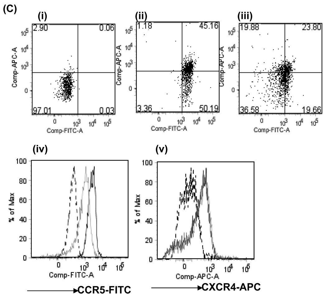
CXCL12 and its receptor CXCR4 play an important role in hematopoietic stem/progenitor cell (HSPC) migration from and retention within the bone marrow. HSPCs are very selective in their chemotactic response and undergo chemotaxis only in response to CXCL12. In addition to CXCR4, HSPCs express receptors for various other chemokines; however, the role of these receptors is not well understood. Freshly isolated CD34(+) cells (highly enriched for HSPCs) from cord blood (CB) express low levels of CCR5; however, if the cells were washed with acidic buffer before Ab staining to remove any ligand bound to CCR5, then nearly 80% of CD34(+) CB cells were found to express CCR5 on the cell surface. Although none of the CCR5 ligands investigated in this study (CCL3, CCL4, and CCL5) induced chemotaxis, at relatively high concentrations they transiently enhanced CXCL12-mediated chemotaxis of CD34(+) CB cells. In contrast, CXCL12-mediated adhesion of cells to VCAM-1-coated surfaces was reduced if CD34(+) CB cells were pretreated with these CCR5 ligands for 15 min. The effect of these chemokines on CXCL12-mediated responses was not at the level of CXCR4 expression, but on downstream signaling pathways elicited by CXCL12. Pretreatment with CCR5 chemokines enhanced CXCL12-mediated Akt phosphorylation, but down-modulated calcium flux in CD34(+) CB cells. Modulation of CXCL12-mediated responses of CD34(+) cells by CCR5 chemokines provides a possible mechanism that underlies movement of HSPCs during inflammation.


Figures

Figure 1
Figure 1. Expression of the CCR5 receptor on CD34+CB cells
(A) CCR5 and CXCR4 expression on CD34+CB was studied by staining freshly isolated unpermeabilized or permeabilized cells with anti-CCR5-FITC (clone 45523) and anti-CXCR4-APC antibodies or the appropriate fluorescent conjugated isotype antibodies and examining the cells using flow cytommetry. (B) Fixed and permeabilized freshly isolated CD34+ CB cells were stained with anti-CCR5-FITC (clone 45523) and anti-CXCR4-APC antibodies and expression of CCR5 and CXCR4 on freshly isolated CD34+CB cells was examined using confocal microscopy. (C) CCR5 and CXCR4 expression on CD34+CB was further studied by staining freshly isolated unpermeabilized or permeabilized cells with a panel of antibodies against various CCR5 epitopes- 1CCR5Ab (clone 45502), 2CCR5Ab (clone 45523), 3CCR5 Ab (clone 45531) and 4CCR5Ab (clone 45549) and anti-CXCR4-APC antibodies. An aliquot of cells was also stained with the appropriate fluorescent conjugated isotype control antibodies and examined using flow cytometry. The data is a representative of 3 independent experiments performed. Shown in the inset are the mean ± SD of mean fluorescence intensity (MFI) of CCR5 expression of three independent experiments.
Figure 2
Figure 2. Effect of CCR5 responsive chemokines on CXCL12 mediated chemotaxis of CD34+CB cells
(A) Freshly isolated CD34+CB cells in 100µl medium containing indicated amount of CCL4 were added to the top well of transwell chamber and CXCL12 (200 ng/ml) was added to the bottom well. Chemotaxis of the cells was assessed 30 mins after initiation of the assay. (B) Effect of CCL4 on CXCL12 mediated chemotaxis of freshly isolated CD34+CB cells was further evaluated by adding CCL4 (1000ng/ml) to the top well alone or to both the top and bottom wells of the transwell chamber. CXCL12 (200ng/ml) was added to the bottom well in each case. Chemotaxis was evaluated 30 mins after initiation of the assay. (C) CCL3, CCL4 or CCL5 (1000 ng/ml) were added to the top well along with freshly isolated CD34+CB cells and CXCL12 (200 ng/ml) was added to the bottom well in a transwell chamber, and chemotaxis of cells was evaluated after 30 mins. (D) Freshly isolated CD34+CB cells along with CCL4 (1000 ng/ml) was added to the top well and chemotaxis to CXCL12 (200ng/ml) was evaluated 30 and 90 mins after initiation of the assay. (E) Chemotaxis of CD34+CB cells in response to various concentration of CXCL12 in the absence or presence of CCL4 (1000ng/ml) was evaluated 30 mins after initiation of chemotaxis assay. The data represents mean ± SD of two experiments performed using CD34+ cells pooled from 3 CB. (F) Freshly isolated CD34+CB cells pretreated with isotype control or anti-CCR5 blocking antibody were added to the top well in the presence of medium or CCL4 (1000ng/ml). In another set of triplicate wells, heat inactivated (HI) CCL5 (1000 ng/ml) was added to the top well. CXCL12 (200 ng/ml) was added to the bottom well in each case. Chemotaxis of the cells was evaluated after 30 mins. The data represents mean ± SD of triplicates of an experiment performed using CD34+ cells pooled from 3 CB. (G) CD34+CB cells was added to the top well in medium alone or in the presence of Met-RANTES (1000 ng/ml) alone, CCL5 (1000 ng/ml) alone or both CCL5 (1000 ng/ml) and Met-RANTES (1000 ng/ml). In the bottom well either medium or CXCL12 (200 ng/ml) was added as indicated. Chemotaxis was measured 30 mins after initiation of experiment. Data represents mean ± SD of triplicates of an experiment performed using CD34+ cells pooled from 3 CB. (H) The percentage of primitive progenitor cells (CD38lo/CD34+) that had migrated under various chemotaxis conditions was determined by staining aliquots of input and migrated cells with anti-CD34-PE and anti-CD38-APC or isotype-matched control antibodies. In all of the above experiments, CD34+ cells isolated from 2–3 CB samples were pooled and used in the assay. The data represents (except for E) mean ±SD of three independent experiments. *p<0.05 compared to chemotaxis in response to CXCL12 alone.
Figure 3
Figure 3. Effect of CCL3, CCL4 and CCL5 on CXCL12 mediated adhesion of CD34+ CB cells to VCAM-1, and on VLA-4 expression
(A) Freshly isolated CD34+CB cells were pretreated with 1000ng/ml of CCL3, CCL4, CCL5 or medium alone for 15 mins and then added to wells pre-coated with VCAM-1 (10µg/ml). CXCL12 or medium was added to the wells and the number of adherent cells was determined after 30 mins. Data represents mean ± SD of three independent experiments. *p<0.05 compared to cells stimulated with CXCL12 alone. (B) Freshly isolated CD34+CB cells were pretreated with CCL4 (1000ng/ml) or medium alone for 15, 30 or 60 mins and then added to wells pre-coated with VCAM-1 (10µg/ml). CXCL12 was added to the wells and the number of adherent cells was determined after 30 mins. Data represents mean ± SD of three independent experiments. *p<0.05 compared to cells stimulated with CXCL12 alone (C) Freshly isolated CD34+CB cells were pretreated with 1000ng/ml of CCL3, CCL4, CCL5 or medium [overlapping lines to the right of isotype control (filled plot)] alone for 15 mins and expression of VLA-4 on these cells was determined by staining the cells with anti-VLA-4-PE antibody and examining the cells using flow cytommeter and analyzing the data using FlowJo software. The data is representative of three independent experiments performed.
Figure 4
Figure 4. Effect of CCL4 on CCR5 and CXCR4 expression on CD34+CB cells
CCR5 and CXCR4 expression on freshly isolated CD34+CB cells when the cells were stained with (Ai) anti-CCR5-FITC (clone 45523) or (Bi) anti-CXCR4-APC antibody without undergoing acid wash prior to antibody staining. Freshly isolated CD34+CB cells were treated with CCL4 (1000ng/ml) for the indicated period of time and then washed once in acidified glycine buffer, followed by a wash in PBS containing 0.2% BSA and then (Aii–iv) stained with anti-CD34P-E antibody along with anti-CCR5-FITC (clone 45523) antibody or (Bii–iv)anti-CXCR4APC antibody. The quadrants were drawn based on staining of cells using appropriate isotype controls. The MFI (mean fluorescence intensity) of CCR5 and CXCR4 expression on CD34+CB cells is shown in each plot shown in upper panel (A). The data is representative of three independent experiments performed. (C) Dot blot presentation of acid washed CD34+CB cells stained with anti-CCR5-FITC (clone 45523) and anti-CXCR4-APC antibodies (Cii) in the absence or (Ciii) presence of CCL4 (100ng/ml). Quadrants were drawn based on staining with (Ci) appropriate isotype controls. Histogram for expression of (Civ) CCR5 and (Cv) CXCR4 on acid washed CD34+ CBcells (black solid line) and in the presence of CCL4 (grey solid line). Isotype staining pattern is shown in the dotted line.
Figure 5
Figure 5. Effect of CCL3, CCL4 and CCL5 on CXCL12 mediated calcium flux, Akt and Erk1/2 phosphorylation, and actin polymerization
(A) Freshly isolated CD34+CB cells were loaded with Fluo-3AM and stimulated with (i) CCL3 (1000 ng/ml) alone (empty bar), CXCL12 (200ng/ml) alone (solid bar), or CCL3 (1000ng/ml) for 120 secs followed by CXCL12 (200ng/ml) (hatched bar) and calcium flux was monitored using flow cytometry and analyzed using FlowJo software. Identical experiments were performed in which instead of CCL3, effects of (ii) CCL4 (1000 ng/ml) and (iii) CCL5 (1000 ng/ml) were evaluated. For each sample described above, baseline fluorescence of Fluo-3AM loaded CD34+CB cells were monitored for 30 secs prior to any stimulation. The data represents mean ± SD of 3 independent experiments. *p<0.05 compared to cells stimulated with CXCL12 alone. (B) Freshly isolated CD34+ cells were pretreated with 1000 ng/ml of CCL3, CCL4, CCL5 or medium alone for 5 mins and then stimulated with CXCL12 for indicated period. At various times post-CXCL12 stimulation, cells were fixed and permeabilized using Cytofix/CytoPerm solution and labeled with phalloidin-Rhodamine for 10 mins at 37°C. Cells were washed with permeabilizing buffer and the amount of F-actin polymerization was examined using flow cytometry, and results were analyzed using Cell Quest software. Data represents mean ± SEM of three independent experiments. *p<0.05 compared to cells stimulated with CXCL12 alone. (C) (i) Freshly isolated CD34+CB cells were kept in IMDM+2%BSA for 1hr. Cells were then pretreated either with medium alone or CCL3 (1000 ng/ml), CCL4 (1000 ng/ml) or CCL5 (1000 ng/ml) for 5 mins. Each sample was thereafter divided in two portions-one portion was washed and lysed and the other portion was stimulated with CXCL12 (200ng/ml) for 3 mins and then washed and lysed. The lysates were run on 4–12% SDS-PAGE and probed with the antibodies. (ii) Densitometric analysis of Akt phosphorylation in CD34+CB cells at baseline (medium alone), following CXCL12 stimulation alone or CCL4 (1000 ng/ml) pretreated CD34+CB cells stimulated with CXCL12 was performed. Ratio of phosphor-Akt to total Akt for each treatment was determined. The data represents mean ± SD of 3 independent experiments. *p<0.05 compared to cells stimulated with CXCL12 alone.
Figure 6
Figure 6. Effect of PI-3kinase inhibitor, LY294002, on chemotaxis and actin polymerization
(A) Freshly isolated CD34+ CB cells were either left untreated or treated with LY294002 (2 µM) for 15 mins. The cells were then added to the top well containing medium or CCL4 (1000 ng/ml). CXCL12 (200 ng/ml) was added to the bottom well. The number of cells that had migrated 30 mins after initiation of the assay were enumerated. Data represents mean ± SD of three experiments. *p<0.05 compared to CD34+CB cells migrated under same conditions but not pretreated with LY294002. (B) Freshly isolated CD34+ CB cells were either left untreated or treated with LY294002 for 15 mins. The cells were thereafter pretreated with CCL4 (1000 ng/ml) for 5 mins and then stimulated with CXCL12 for indicated time period. Following treatment for the indicated time periods, cells were fixed and permeabilized using Cytofix/CytoPerm solution and labeled with phalloidin-Rhodamine for 10 mins at 37°C. The cells were washed with permeabilizing buffer, the amount of F-actin polymerization was examined using flow cytometry, and the results were analyzed using Cell Quest software. Data represents mean ± SD of three experiments. *p<0.05 compared to CD34+CB cells not treated with LY294002.
Figure 7
Figure 7. Proposed model for CCR5-ligand modulation of CXCL12 mediated responses
(A) CXCL12 binding to its receptors CXCR4 results in activation of various signaling pathways including calcium mobilization and activation of Erk and Akt. Activation of Erk and Akt play a role in CXCL12-mediated chemotaxis (solid line). Calcium mobilization in response to G-PCR activation is known to enhance adhesion, although its role specifically in CXCL12 mediated adhesion of CD34+ cells is not defined (dashed line). (B) In the presence of CCR5 ligands, CXCL12-mediated Akt phosphorylation is enhanced (thick arrow) and is accompanied by a transient increase in chemotaxis of CD34+CB cells towards CXCL12. However, CCR5 ligands down modulate CXCL12 mediated calcium flux (thin arrow) and also CXCL12 mediated adhesion.

Similar articles

Cited by

References

    1. Ma Q, Jones D, Borghesani PR, Segal RA, Nagasawa T, Kishimoto T, Bronson RT, Springer TA. Impaired B-lymphopoiesis, myelopoiesis, and derailed cerebellar neuron migration in C. Proc. Natl. Acad. Sci. U. S. A. 1998;95:9448–9453. - PMC - PubMed
    1. Zou YR, Kottmann AH, Kuroda M, Taniuchi I, Littman DR. Function of the chemokine receptor CXCR4 in haematopoiesis and in cerebellar development. Nature. 1998;393:595–599. - PubMed
    1. Levesque JP, Hendy J, Takamatsu Y, Simmons PJ, Bendall LJ. Disruption of the CXCR4/CXCL12 chemotactic interaction during hematopoietic stem cell mobilization induced by GCSF or cyclophosphamide. J. Clin. Invest. 2003;111:187–196. - PMC - PubMed
    1. Broxmeyer HE, Orschell CM, Clapp DW, Hangoc G, Cooper S, Plett PA, Liles WC, Li X, Graham-Evans B, Campbell TB, Calandra G, Bridger G, Dale DC, Srour EF. Rapid mobilization of murine and human hematopoietic stem and progenitor cells with AMD3100, a CXCR4 antagonist. J. Exp. Med. 2005;201:1307–1318. - PMC - PubMed
    1. Aiuti A, Webb IJ, Bleul C, Springer T, Gutierrez-Ramos JC. The chemokine SDF-1 is a chemoattractant for human CD34+ hematopoietic progenitor cells and provides a new mechanism to explain the mobilization of CD34+ progenitors to peripheral blood. J. Exp. Med. 1997;185:111–120. - PMC - PubMed

Publication types

MeSH terms