. 2009 Dec;20(12):2604-15.
doi: 10.1681/ASN.2009010122. Epub 2009 Nov 16.

Tracing the origin of glomerular extracapillary lesions from parietal epithelial cells

Affiliations

Tracing the origin of glomerular extracapillary lesions from parietal epithelial cells

Bart Smeets et al. J Am Soc Nephrol. 2009 Dec.

Abstract

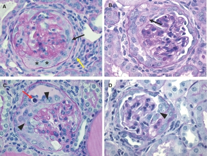
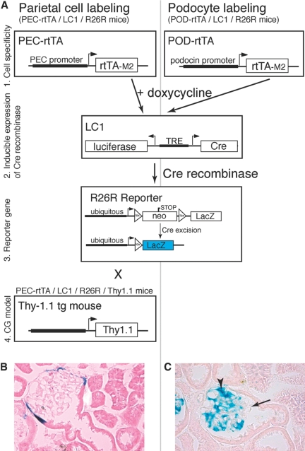
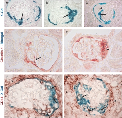
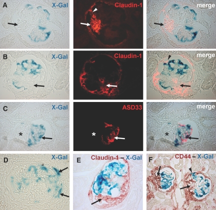
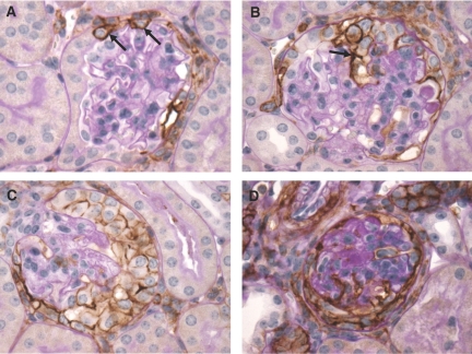
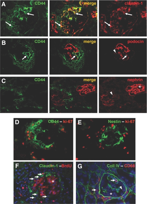
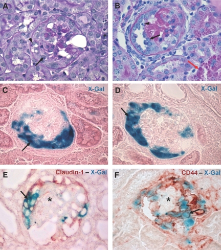
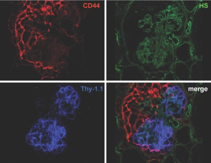
Cellular lesions form in Bowman's space in both crescentic glomerulonephritis and collapsing glomerulopathy. The pathomechanism and origin of the proliferating cells in these lesions are unknown. In this study, we examined proliferating cells by lineage tracing of either podocytes or parietal epithelial cells (PECs) in the nephrotoxic nephritis model of inflammatory crescentic glomerulonephritis. In addition, we traced the fate of genetically labeled PECs in the Thy-1.1 transgenic mouse model of collapsing glomerulopathy. In both models, cellular bridges composed of PECs were observed between Bowman's capsule and the glomerular tuft. Genetically labeled PECs also populated larger, more advanced cellular lesions. In these lesions, we detected de novo expression of CD44 in activated PECs. In contrast, we rarely identified genetically labeled podocytes within the cellular lesions of crescentic glomerulonephritis. In conclusion, PECs constitute the majority of cells that compose early extracapillary proliferative lesions in both crescentic glomerulonephritis and collapsing glomerulopathy, suggesting similar pathomechanisms in both diseases.


Figures

Figure 1.
Figure 1.
Histology of the CrGN model. (A–D) PAS stainings of the CrGN model at day 14 after anti-nephrotoxic serum injection. Light microscopy revealed pronounced hyperplasia of glomerular epithelial cells, forming organized multilayered true crescentic lesions (A and B, arrows) and less organized pseudocrescents and monolayer lesions on the glomerular tuft (C and D, arrowheads). Marked periglomerular fibrosis was seen in the regions surrounding affected glomeruli (A, yellow arrow). An occasional infiltrative inflammatory cell (C, red arrow, polymorphonuclear leukocyte) or protein accumulation (A, asterisks) could be observed within the cellular lesions.
Figure 2.
Figure 2.
Genetic tagging of podocytes or PECs in triple transgenic doxycycline-inducible mouse lines. (A) The modified rabbit podocalyxin promoter (PEC promoter) drives expression of the enhanced tetracycline reverse transactivator (rtTA-M2) as described previously. The podocyte-specific Pod-rtTA transgenic mice were kindly provided by Jeffrey Kopp, Massachusetts Institute of Technology, Cambridge, MA. Both mice were mated with LC1 mice expressing Cre recombinase (and luciferase) under the control of tetracycline responsive elements (TREs), which can be activated by the transcription factor rtTA-M2 in the presence of doxycycline. The R26R mice were used as a reporter line, which irreversibly expresses β-galactosidase (LacZ) under the control of the ubiquitously active Rosa26 locus only after Cre excision of an interposed floxed neomycin cassette (neo, acting as a stop signal) has occurred. These matings resulted in the triple transgenic PEC-rtTA/LC1/R26R (to tag parietal cells) and POD-rtTA/LC1/R26R mice (to tag podocytes). For studies in the CG model, the PEC-rtTA/LC1/R26R mice were mated to the Thy-1.1 transgenic mouse line (T6 clone). (B) Genetic labeling of PECs using doxycycline at the age of 4 wk in triple transgenic PEC-rtTA/LC1/R26R mice. After induction, Cre recombination occurred in more than 70% of all PECs (arrowheads, labeled PEC). Sporadic labeling was observed in some tubular cells of the renal cortex (not shown). (C) Genetic labeling of podocytes using doxycycline at the age of 4 wk in triple transgenic Pod-rtTA/LC1/R26R mice. After induction, Cre recombination occurred in more than 70% of all podocytes, similar to the PEC-rtTA mice (arrowheads, labeled podocytes; arrow, unlabeled tuft segment).
Figure 3.
Figure 3.
Genetically labeled PECs are present within crescentic lesions. (A–C) The PECs that were tagged genetically after doxycycline administration in PEC-rtTA/LC1/R26R mice were traced in crescentic lesions. True crescentic lesions (A), pseudocrescents (B), and monolayer lesions (C) observed at day 14 after the NTS injection stained positive with X-gal, confirming their origin from the parietal epithelial compartment (arrows). A cross section of the glomerular tuft is seen separating the monolayer lesion and Bowman's capsule in C (arrowhead). (D) β-Gal expressing cells (stained using Bluo-Gal, resulting in intracellular blue particles) frequently formed cell bridges between Bowman's capsule and the glomerular tuft (D, arrow). The Bluo-gal expressing cells showed colocalization with the PEC marker claudin-1 (D, arrow; E, arrowhead). Within monolayer (F, arrow) and crescentic lesions (G, arrows), the X-gal positive cells stained for CD44. Note that CD44 also is expressed by the cells present in the periglomerular fibrotic region (F and G, arrowheads).
Figure 4.
Figure 4.
Genetically labeled podocytes are excluded from crescentic lesions. (A, B) Double staining with X-gal and claudin-1 revealed no costaining in the β-gal expressing cells. Genetic labeling persisted in podocytes. But the crescentic lesions in Bowman's space that stain positive for claudin-1 were X-gal negative (A and B, arrows). Of note is the finding that the typical visceral hyperplastic lesion positive for claudin-1 is connected to the claudin-1 positive cells lining Bowman's capsule (A, arrowhead). (C) X-gal and ASD33 immunofluorescence double staining showing costaining β-gal expressing podocytes with the podocyte marker antibody ASD33. The affected segment of the glomerulus is negative for X-gal and ASD33 (C, asterisks). The merged images show a pseudocolor overlay generated with Adobe Photoshop 7.0 (Adobe Systems Incorporated, San Jose, CA). (D) X-gal staining showing rare X-gal positive cells that are present along the periphery of glomerular lesions (D, arrows). (E, F) Immunohistochemical double staining for claudin-1 and CD44 with X-gal revealed no costaining. The crescentic lesions were positive for claudin-1 and CD44 but generally negative for X-gal (E and F, arrows). Occasionally, involvement of podocytes within the lesion was observed (F, arrowhead). Interestingly, in this particular lesion the podocyte seems to be attached to Bowman's capsule, a rare finding.
Figure 5.
Figure 5.
PAS–CD44 double staining. (A) A still morphologically normal glomerulus in the CrGN mouse that shows CD44 expression by PECs on Bowman's capsule (A, arrows). (B) A glomerulus containing a small pseudocrescent, which stains for CD44 (B, arrow). (C) Large crescentic lesion showing a homogenous CD44 positive cell population in Bowman's space. (D) True crescentic lesion with flattened cells that circumvent the glomerular tuft. The cells forming the crescent stain for CD44.
Figure 6.
Figure 6.
Phenotype of proliferating cells (A) Double immunofluorescence staining for claudin-1 (red) and CD44 (green). CD44 is de novo expressed by the cells forming the crescentic lesions. Outside of the glomerulus, CD44 is expressed by a subpopulation of the cells within the periglomerular fibrotic area and some tubular cells. Within the crescentic lesions, the CD44 positive cells costain with claudin-1 (A, arrows). (B, C) Double immunofluorescence staining for CD44 (green) and the podocyte markers podocin (B) and nephrin (C). The proliferating cells filling up Bowman's space are positive for CD44 and negative for the podocyte markers. Note that the CD44 positive cells are positioned directly against the podocin positive podocytes (B, arrow). The glomerular tufts of affected glomeruli showed segmental (B, arrowhead) to a global (C, arrowhead) reduction in podocyte marker staining. Panel C also shows a morphologically normal glomerulus showing no CD44 staining and a normal nephrin staining (C, asterisks). (D) Double immunofluorescence staining for CD44 (green) and the proliferation marker Ki-67 (red). Many CD44 positive cells in the periglomerular area and within the crescentic lesions express the proliferation marker Ki-67. (E) Double immunofluorescence staining for nestin (green) and Ki-67 (red). Only rare nestin positive cells show costaining with the proliferation marker Ki-67. The majority of the Ki-67 positive cells were detected in the area peripheral to the nestin positive cells. (F) Double immunofluorescence for the proliferation marker BrdU (red) and claudin-1 (green). The nuclei are counterstained with Hoechst to reveal nuclei (blue). Many claudin-1 positive cells forming the cellular lesion stain positive for BrdU. Together with the Hoechst staining, BrdU positive cells show purple nuclei (arrows). The secondary anti-mouse antibody used for the detection of the BrdU antibody also recognized the endogenous mouse antibodies that bind to the GBM (F, asterisks). (G) Double immunofluorescence staining for collagen IV (green) and the macrophage marker CD68 (red). The nuclei are counterstained with Hoechst (blue). The cells forming the crescentic lesion do not express CD68 (G, asterisks). Many CD68 positive cells were detected in the periglomerular areas surrounding affected glomeruli and within the glomerular tuft (G, arrows).
Figure 7.
Figure 7.
Genetically labeled PECs are present within the CG lesions. (A, B) PAS staining of kidney sections at day 7 after the anti-Thy-1.1 mAb injection. The early lesions in the PEC-rtTA/Thy-1.1 mice showed segmental to global collapse of the glomerular tuft, hypertrophy and hyperplasia of the glomerular epithelial cells that resulted in the formation of pseudocrescents (A, arrow), and monolayer lesions on the glomerular tuft (B, arrow). The arrowhead in panel A shows vacuoles and resorption droplets, a sign of podocyte injury. The arrowhead in panel B shows a mitotic cell body in a monolayer lesion. Protein casts were observed frequently in the tubular structures. In addition, many proximal tubular cells contained protein resorption droplets (B, red arrow). (C, D) The CG lesions stained positive with X-gal (C and D, arrows). (E, F) Double staining of X-gal with claudin-1 and CD44, respectively. Within cellular lesions in Bowman's space, X-gal positive cells costain with claudin-1 (E, arrow) and CD44 (F, arrow), whereas the cells of the glomerular tuft remain negative for the PEC markers (E and F, asterisks).
Figure 8.
Figure 8.
Triple immunofluorescence staining for CD44, Thy-1.1, and heparan sulfates (HS). The HS panel, in which the glomerular basal membranes are stained with an antibody directed against heparan sulfates, shows a glomerulus with a collapsed glomerular tuft. The epithelial cells in Bowman's space show expression of CD44. Only at the margin of the glomerular tuft we sometimes observed colocalization of CD44 and the Thy-1.1 antigen expressed by podocytes, indicating that the Thy1.1 positive podocytes are covered directly with the CD44 positive cells of the lesion.

References

    1. Jennette JC, Olson JL, Schwartz MM, Silva FG: Crescentic glomerulonephritis. In: Heptinstall's Pathology of the Kidney, 6th Ed., Philadelphia, Lippincott-Raven, pp 615–641, 2005
    1. Le Hir M, Besse-Eschmann V: A novel mechanism of nephron loss in a murine model of crescentic glomerulonephritis. Kidney Int 63: 591–599, 2003 - PubMed
    1. Detwiler RK, Falk RJ, Hogan SL, Jennette JC: Collapsing glomerulopathy: A clinically and pathologically distinct variant of focal segmental glomerulosclerosis. Kidney Int 45: 1416–1424, 1994 - PubMed
    1. D'Agati V, Suh JI, Carbone L, Cheng JT, Appel G: Pathology of HIV-associated nephropathy: A detailed morphologic and comparative study. Kidney Int 35: 1358–1370, 1989 - PubMed
    1. Moudgil A, Nast CC, Bagga A, Wei L, Nurmamet A, Cohen AH, Jordan SC, Toyoda M: Association of parvovirus B19 infection with idiopathic collapsing glomerulopathy. Kidney Int 59: 2126–2133, 2001 - PubMed

Publication types

LinkOut - more resources