

Lack of glutathione peroxidase 1 accelerates cardiac-specific hypertrophy and dysfunction in angiotensin II hypertension
- PMID: 19917877
- PMCID: PMC3061336
- DOI: 10.1161/HYPERTENSIONAHA.109.135715
Lack of glutathione peroxidase 1 accelerates cardiac-specific hypertrophy and dysfunction in angiotensin II hypertension
Abstract
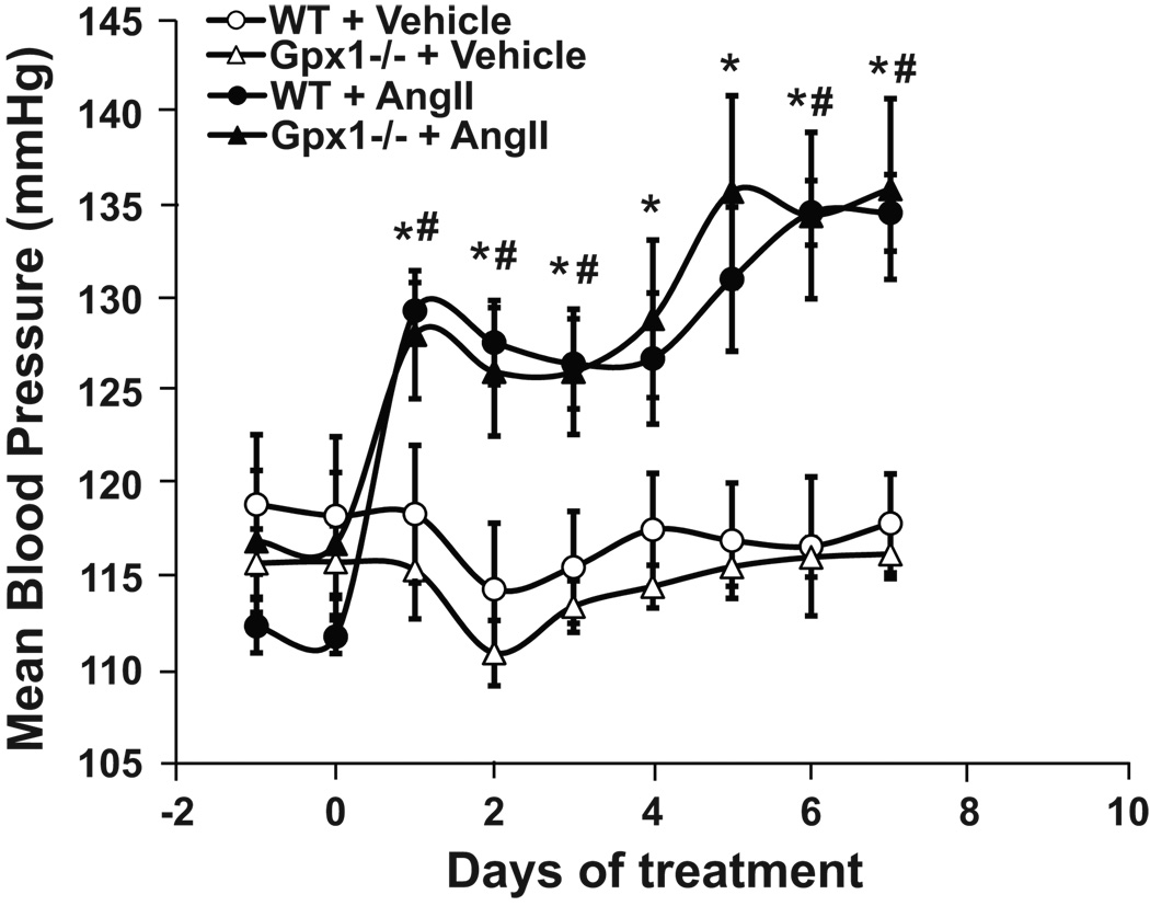
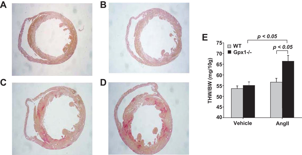
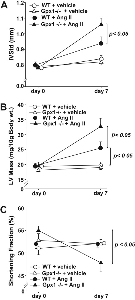
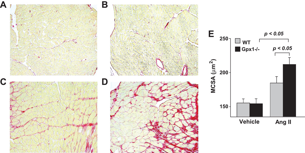
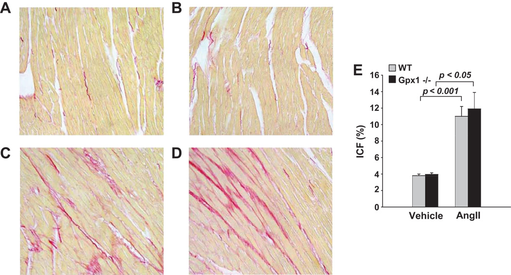
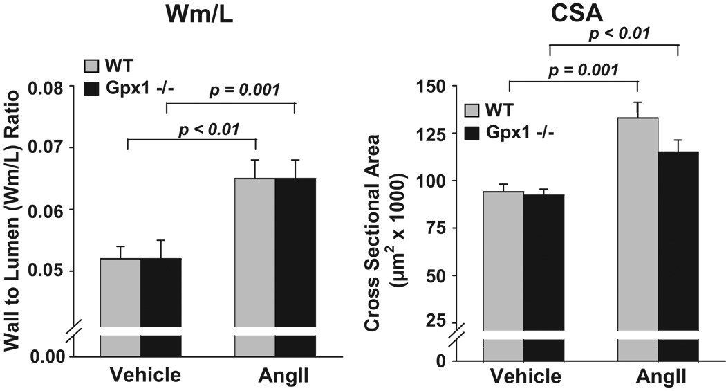
Glutathione peroxidase 1 (Gpx1) plays an important role in cellular defense by converting hydrogen peroxide and organic hydroperoxides to nonreactive products, and Gpx1(-/-) mice, which are characterized by reduced tissue glutathione peroxidase activity, are known to exhibit enhanced oxidative stress. Peroxides participate in tissue injury, as well as the hypertrophy of cultured cells, yet the role of Gpx1 to prevent end organ damage in cardiovascular tissue is not clear. We postulated that Gpx1 deletion would potentiate both aortic and cardiac hypertrophy, as well as mean arterial blood pressure, in response to angiotensin II (AngII). Our results show that short-term AngII markedly increased left ventricular mass, myocyte cross-sectional area, and interventricular septum thickness and decreased shortening fraction in Gpx1(-/-) mice as compared with wild-type animals. On the other hand, AngII resulted in a similar increase in mean arterial blood pressure in wild-type and Gpx1(-/-) mice. Collagen deposition increased in response to AngII, but no differences were found between strains. Vascular hypertrophy increased to the same extent in Gpx1(-/-) and wild-type mice. Collectively, our results indicate that Gpx1 deficiency accelerates cardiac hypertrophy and dysfunction but has no effect on vascular hypertrophy and mean arterial blood pressure and suggest a major role for Gpx1 in cardiac dysfunction in AngII-dependent hypertension.
Figures
References
-
- Gardin JM, McClelland R, Kitzman D, Lima JA, Bommer W, Klopfenstein HS, Wong ND, Smith VE, Gottdiener J. M-mode echocardiographic predictors of six- to seven-year incidence of coronary heart disease, stroke, congestive heart failure, and mortality in an elderly cohort (the Cardiovascular Health Study) Am J Cardiol. 2001;87:1051–1057. - PubMed
-
- Verdecchia P, Porcellati C, Reboldi G, Gattobigio R, Borgioni C, Pearson TA, Ambrosio G. Left ventricular hypertrophy as an independent predictor of acute cerebrovascular events in essential hypertension. Circulation. 2001;104:2039–2044. - PubMed
-
- Frohlich ED. Overview of hemodynamic and non-hemodynamic factors associated with left ventricular hypertrophy. J Mol Cell Cardiol. 1989;21:3–10. - PubMed
-
- Hanevold C, Waller J, Daniels S, Portman R, Sorof J. The effects of obesity, gender, and ethnic group on left ventricular hypertrophy and geometry in hypertensive children: a collaborative study of the International Pediatric Hypertension Association. Pediatrics. 2004;113:328–333. - PubMed
-
- Cuspidi C, Muiesan ML, Valagussa L, Salvetti M, Di Biagio C, Agabiti-Rosei E, Magnani B, Zanchetti A. Comparative effects of candesartan and enalapril on left ventricular hypertrophy in patients with essential hypertension: the candesartan assessment in the treatment of cardiac hypertrophy (CATCH) study. J Hypertens. 2002;20:2293–2300. - PubMed
Publication types
MeSH terms
Substances
Grants and funding
LinkOut - more resources
Full Text Sources
Medical
Molecular Biology Databases
Miscellaneous
