. 2009 Dec 1;106(48):20423-8.
doi: 10.1073/pnas.0910371106. Epub 2009 Nov 16.

Epidermal loss of JunB leads to a SLE phenotype due to hyper IL-6 signaling

Affiliations

Epidermal loss of JunB leads to a SLE phenotype due to hyper IL-6 signaling

Pamina Pflegerl et al. Proc Natl Acad Sci U S A. .

Abstract

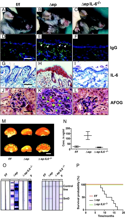
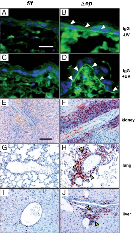
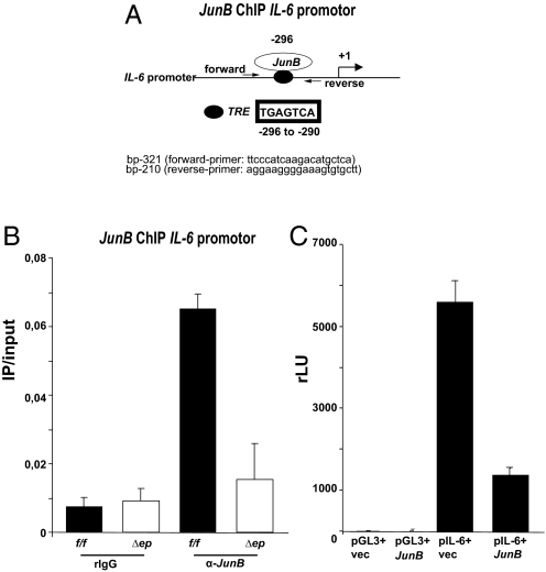
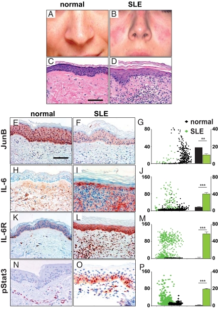
Systemic lupus erythematosus (SLE) is a complex autoimmune disease affecting various tissues. Involvement of B and T cells as well as increased cytokine levels have been associated with disease manifestation. Recently, we demonstrated that mice with epidermal loss of JunB (JunB(Deltaep)) develop a myeloproliferative syndrome (MPS) due to high levels of G-CSF which are secreted by JunB-deficient keratinocytes. In addition, we show that JunB(Deltaep) mice develop a SLE phenotype linked to increased epidermal interleukin 6 (IL-6) secretion. Intercrosses with IL-6-deficient mice could rescue the SLE phenotype. Furthermore, we show that JunB binds to the IL-6 promoter and transcriptionally suppresses IL-6. Facial skin biopsies of human SLE patients similarly revealed low JunB protein expression and high IL-6, activated Stat3, Socs-1, and Socs-3 levels within lupus lesions. Thus, keratinocyte-induced IL-6 secretion can cause SLE and systemic autoimmunity. Our results support trials to use alpha-IL-6 receptor antibody therapy for treatment of SLE.


Conflict of interest statement

The authors declare no conflict of interest.

Figures

Fig. 1.
Fig. 1.
SLE-like skin and kidney affections in JunBΔep mice. A 3-month-old wild-type mouse with normal skin was compared to a JunBΔep littermate control (A) with ulcerative skin lesions in the face region (B) and rescue of the skin phenotype after crossing to IL-6−/− mice (C). In indirect immunofluorescence using α-IgG (D–F), JunBΔep mice (E) (arrowheads) show a prominent “lupus band” in the epidermal-dermal junction, whereas in wild-type (D) and JunBΔepIL-6−/− mice (F), this band is absent. (Scale bar, 50 μm.) The skin of wild-type and JunBΔepIL-6−/− mice show no IL-6 expression, whereas JunBΔep mice have high IL-6 expression levels (G–I). In the AFOG staining, the glomerular changes are characterized by mesangial hypercellularity with glomerular basement membrane thickening and luminal obstruction by hyaline immune complex deposits in the mutant (arrowheads) (K) compared to wild-type (J) and JunBΔepIL-6−/− (L). (Scale bar, 50 μm.) The macroscopic kidney appearance (M) of 6-month-old wild-type mice compared to small and atrophic kidneys of mutant mice. The kidney size appears to be normal in the JunBΔepIL-6−/− intercross. (Scale bar, 0.3 cm.) Note the normal width of the kidney cortex in wild-type and JunBΔepIL-6−/− mice (arrowheads) compared to the mutant. Albumin ELISA shows increased protein levels in the urine of JunBΔep mice compared to JunBf/f controls and JunBΔepIL-6−/− (N). Immunoblot analysis (O) showing increased anti-histone antibody serum levels against the individual histones H1, H2A, H2B, H3, and H4 in JunBΔep mice: Lanes 1 and 2, 6-month-old JunBf/f mice; lanes 3 and 4, 6-month-old JunBΔep mice with high α-histone Ab reactivities similar to the patient in lane 7. Lanes 5 and 6, 6-month-old JunBΔepIL-6−/− mice. Lane 7, SLE patient serum with high α-histone Ab reactivities. JunBΔep mice show increased titers of α-histone Abs with cumulative age. No anti-histone antibodies can be found in JunBΔepIL-6−/− mice. Line immunoassays using sera of mice were performed. JunBΔep mice show pronounced α-SmD Abs (lanes 2 and 3), similar to a SLE patient serum (lane 6). No α-SmD antibodies are found in sera of JunBf/f wild-type mice (lane 1) and JunBΔepIL-6−/− mice (lanes 4 and 5). (P) The survival time of JunBΔep mice was reduced compared to wild-type mice (Kaplan Meier blot).
Fig. 2.
Fig. 2.
UV irradiation experiments of JunBf/f and JunBΔep mice (A–D). (Scale bar, 50 μm.) JunBf/f show no α-IgG immunofluorescence with or without UV (A and C). JunBΔep mice show linear α-IgG-immunofluorescence at the dermo-epidermal junction (arrowheads) (B), which becomes more intense after UV-irradiation (D). Thus, UV-irradiation enhanced severeness of lupus-like skin lesions in JunBΔep mice on immunofluorescence levels. JunBΔep mice show dense lymphocytic infiltrates in the peripheral organs kidney, lung, and liver compared to JunBf/f control mice. These infiltrates are mainly composed of αCD3 positive T-cells (E–J) (arrowheads). (Scale bar, 100 μm.)
Fig. 3.
Fig. 3.
Schematic overview of known transcription factor binding sites in the IL-6 promoter, indicating the location of the putative AP-1 site (filled circle) and the transcription start site (+1) (A). Arrows indicate the position of the primers used for ChIP. ChIP using an JunB Ab or IgG control Ab reveals binding of JunB to the IL-6 promoter in a region containing a conserved AP-1 binding site in keratinocytes isolated from JunBfl/fl mice. No binding was detected in JunBΔep keratinocytes (B). HeLa cells were either cotransfected with pCDNA4_hismax_JUNB (JunB) or pCDNA4_hismaxA (vec) together with either pIL-6 or pGL3 empty vector for control purpose. Twenty-four hours later, luciferase- and β-gal activity (expressed from a routinely cotransfected β-gal expression vector) was measured. Luciferase expression from two replicates, normalized to β-gal expression was plotted, and error bars are given, represent ± SD. Relative normalized luciferase expression was below detection limit from promoterless pGL3-basic plasmid, while intact luciferase expression from pIL-6 was abrogated by ectopic JunB expression from pCDNA4_hismax_JUNB (C).
Fig. 4.
Fig. 4.
Normal human facial skin (A) compared to SLE patient's skin (B). SLE combines systemic with cutaneous lesions such as the “butterfly” facial lesion, resembling the cardinal feature of human SLE. Compared to normal human skin (C), histology of the patient's skin (D) revealed typical vacuolar alterations of the basal cell layers, a predominant basal membrane, vasculitis of dermal blood vessels, and dermal chronic inflammatory infiltrates consisting mainly of lymphocytes. (Scale bar, 100 μm.) JunB expression is high in normal facial skin (E) when compared to the reduced JunB expression levels in SLE patient's facial skin area (F), which was quantified by automated cell acquisition and quantification software (Histoquest) (G). Immunohistochemistry of SLE patient's skin revealed high protein expression levels of IL-6 (I), IL-6R (L), and pStat3 (O) especially in the basal cell layers, compared to normal facial skin biopsies (H, K, and N, respectively). (Scale bar, 50 μm.) On the right side, the respective protein quantification results of the human patient samples depicted in (J, M, P) using Histoquest software are shown as scattergrams and bar diagrams. The relative protein expression levels compare normal (black dots/bars) versus SLE patients (green dots/bars).

Similar articles

Cited by

References

    1. Kishimoto T. Interleukin-6: From basic science to medicine—40 years in immunology. Annu Rev Immunol. 2005;23:1–21. - PubMed
    1. Heinrich PC, et al. Principles of interleukin (IL)-6-type cytokine signalling and its regulation. Biochem J. 2003;374:1–20. - PMC - PubMed
    1. Hirano T. Interleukin 6 and its receptor: Ten years later. Int Rev Immunol. 1998;16:249–284. - PubMed
    1. Youinou P, Jamin C. The weight of interleukin-6 in B cell-related autoimmune disorders. J Autoimmun. 2009;32:206–210. - PubMed
    1. Eitner F, et al. Role of interleukin-6 in mediating mesangial cell proliferation and matrix production in vivo. Kidney Int. 1997;51:69–78. - PubMed

Publication types

MeSH terms