. 2009 Dec 1;106(48):20504-9.
doi: 10.1073/pnas.0908083106. Epub 2009 Nov 16.

GluN2B subunit-containing NMDA receptor antagonists prevent Abeta-mediated synaptic plasticity disruption in vivo

Affiliations

GluN2B subunit-containing NMDA receptor antagonists prevent Abeta-mediated synaptic plasticity disruption in vivo

Neng-Wei Hu et al. Proc Natl Acad Sci U S A. .

Erratum in

  • Proc Natl Acad Sci U S A. 2010 Jul 13;107(28):12734. Anwy, Roger [corrected to Anwyl, Roger]

Abstract

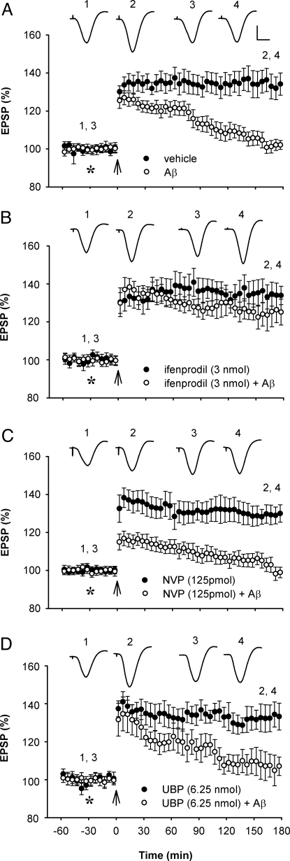
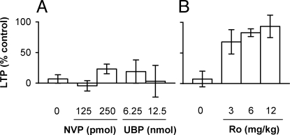
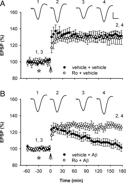
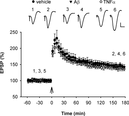
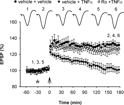
Currently, treatment with the relatively low-affinity NMDA receptor antagonist memantine provides limited benefit in Alzheimer's disease (AD). One probable dose-limiting factor in the use of memantine is the inhibition of NMDA receptor-dependent synaptic plasticity mechanisms believed to underlie certain forms of memory. Moreover, amyloid-beta protein (Abeta) oligomers that are implicated in causing the cognitive deficits of AD potently inhibit this form of plasticity. Here we examined if subtype-preferring NMDA receptor antagonists could preferentially protect against the inhibition of NMDA receptor-dependent plasticity of excitatory synaptic transmission by Abeta in the hippocampus in vivo. Using doses that did not affect control plasticity, antagonists selective for NMDA receptors containing GluN2B but not other GluN2 subunits prevented Abeta(1-42) -mediated inhibition of plasticity. Evidence that the proinflammatory cytokine TNFalpha mediates this deleterious action of Ass was provided by the ability of TNFalpha antagonists to prevent Abeta(1-42) inhibition of plasticity and the abrogation of a similar disruptive effect of TNFalpha using a GluN2B-selective antagonist. Moreover, at nearby synapses that were resistant to the inhibitory effect of TNFalpha, Abeta(1-42) did not significantly affect plasticity. These findings suggest that preferentially targeting GluN2B subunit-containing NMDARs may provide an effective means of preventing cognitive deficits in early Alzheimer's disease.


Conflict of interest statement

The authors declare no conflict of interest.

Figures

Fig. 1.
Fig. 1.
Low-dose NMDAR antagonist selective for GluN2B but not GluN2A or GluN2C/D subunits abrogates Aβ1–42-mediated inhibition of LTP in vivo. (A) Intracerebroventricular (i.c.v., asterisk) injection of soluble Aβ1–42 (80 pmol) inhibited high frequency stimulation (arrow) -induced LTP (n = 6; P < 0.05 compared with vehicle, n = 6; P > 0.05 compared with baseline; two-way ANOVA with repeated measures and paired t tests). (B) A low dose (3 nmol, i.c.v.) of the GluN2B selective antagonist ifenprodil that did not affect LTP on its own (n = 5), prevented the inhibition of LTP by Aβ1–42 (n = 6; P < 0.05 compared with Aβ1–42 alone). (C) A relatively low dose (125 pmol, i.c.v.) of the GluN2A selective antagonist NVP-AAM077 that did not affect LTP on its own (n = 5), failed to prevent the inhibition of LTP by Aβ1–42 (n = 6; P > 0.05). (D) Similarly, a relatively low dose (6.25 nmol, i.c.v.) of the GluN2C/D selective antagonist UBP141 that did not affect LTP on its own (n = 4), failed to prevent the inhibition of LTP by Aβ1–42 (n = 4; P > 0.05). Values are the mean percentage of pre-HFS baseline EPSP amplitude (±SEM). Insets show representative EPSP traces at the times indicated. Calibration bars: vertical, 2 mV; horizontal, 10 ms.
Fig. 2.
Fig. 2.
Dose-dependence of the effects of subtype-selective NMDAR antagonists on the inhbition of LTP by Aβ1–42. (A) Neither pretreatment with the GluN2A antagonist NVP-AAM077 (125 pmol, n = 5; and 250 pmol, n = 4, i.c.v.) nor the GluN2C/D antagonist UBP141 (6.25, n = 4; and 12.5 pmol, n = 4, i.c.v.) significantly affected the inhibition of LTP by Aβ1–42 (80 pmol, i.c.v., n = 6 for Aβ1–42 alone) (P > 0.05, one-way ANOVA). (B) In contrast, pretreatment with the GluN2B antagonist Ro 25–6981 (3 mg/kg, n = 4; 6 mg/kg, n = 6; and 12 mg/kg, n = 4, i.p.) significantly (P < 0.05) reduced the Aβ1–42-mediated inhibition of LTP (n = 7 for Aβ1–42 alone). LTP values are expressed as the mean (±SEM) % control magnitude of LTP at 3 h after high frequency conditioning stimulation.
Fig. 3.
Fig. 3.
Systemic treatment with the GluN2B subunit-selective NMDAR antagonist Ro 25–6981 prevents Aβ1–42-mediated inhibition of LTP. (A) Systemic administration of Ro 25–6981 (6 mg/kg, i.p.) did not significantly affect LTP (n = 5; P > 0.05 compared with vehicle-injected controls, n = 5). (B) Pretreatment with Ro 25–6981 prevented the inhibition of LTP caused by Aß1–42 (80 pmol, i.c.v., asterisk) (n = 7; P < 0.05 compared with Aβ1–42 alone, n = 6). Values are the mean percentage of pre-HFS baseline EPSP amplitude (±SEM). Calibration bars for EPSP traces: vertical, 2 mV; horizontal, 10 ms.
Fig. 4.
Fig. 4.
Resistance of LTP induction at basal dendrites to the inhibitory effect of TNFα and Aβ1–42. High frequency stimulation (arrows) induced robust LTP of synaptic transmission at basal dendrites of CA1 pyramidal cells in the stratum oriens of animals injected i.c.v. with either vehicle (5 μL, n = 6, P < 0.05) (closed circles), TNFα (1.5 pmol, n = 6, P < 0.05) (open circles) or Aß1–42 (320 pmol, n = 6, P < 0.05) (triangles). Values are the mean percentage of pre-HFS baseline EPSP amplitude (±SEM). Calibration bars for EPSP traces: vertical, 0.5 mV; horizontal, 10 ms.
Fig. 5.
Fig. 5.
Systemic treatment with the GluN2B subunit-selective NMDAR antagonist Ro 25–6981 prevents TNFα-mediated inhibition of LTP. Pretreatment with Ro 25–6981 prevented the inhibition of LTP caused by TNFα (1.5 pmol, i.c.v.) (n = 6; P < 0.05 compared with TNFα alone, n = 5; P > 0.05 compared with vehicle-injected controls, n = 5). Values are the mean percentage of pre-HFS baseline EPSP amplitude (±SEM). Calibration bars for EPSP traces: see Fig. 1.

Similar articles

Cited by

References

    1. Hynd MR, Scott HL, Dodd PR. Glutamate-mediated excitotoxicity and neurodegeneration in Alzheimer's disease. Neurochem Int. 2004;45:583–595. - PubMed
    1. Haass C, Selkoe DJ. Soluble protein oligomers in neurodegeneration: Lessons from the Alzheimer's amyloid beta-peptide. Nat Rev Mol Cell Biol. 2007;8:101–112. - PubMed
    1. Lambert MP, et al. Diffusible, nonfibrillar ligands derived from A beta(1–42) are potent central nervous system neurotoxins. Proc Natl Acad Sci USA. 1998;95:6448–6453. - PMC - PubMed
    1. Shankar GM, et al. Amyloid-beta protein dimers isolated directly from Alzheimer's brains impair synaptic plasticity and memory. Nat Med. 2008;14:837–842. - PMC - PubMed
    1. Raina P, et al. Effectiveness of cholinesterase inhibitors and memantine for treating dementia: Evidence review for a clinical practice guideline. Ann Intern Med. 2008;148:379–397. - PubMed

Publication types

MeSH terms