. 2009 Nov 24;106(47):19940-5.
doi: 10.1073/pnas.0907898106. Epub 2009 Nov 16.

Adenovirus vector vaccination induces expansion of memory CD4 T cells with a mucosal homing phenotype that are readily susceptible to HIV-1

Affiliations

Adenovirus vector vaccination induces expansion of memory CD4 T cells with a mucosal homing phenotype that are readily susceptible to HIV-1

Adel Benlahrech et al. Proc Natl Acad Sci U S A. .

Abstract

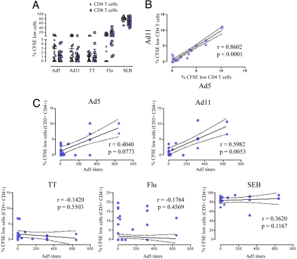
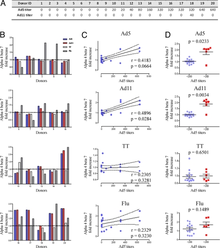
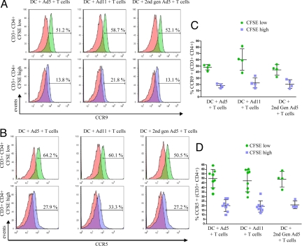
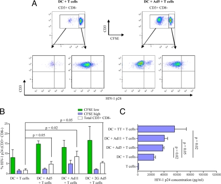
In the recently halted HIV type 1 (HIV-1) vaccine STEP trial, individuals that were seropositive for adenovirus serotype 5 (Ad5) showed increased rates of HIV-1 infection on vaccination with an Ad5 vaccine. We propose that this was due to activation and expansion of Ad5-specific mucosal-homing memory CD4 T cells. To test this hypothesis, Ad5 and Ad11 antibody titers were measured in 20 healthy volunteers. Dendritic cells (DCs) from these individuals were pulsed with replication defective Ad5 or Ad11 and co-cultured with autologous lymphocytes. Cytokine profiles, proliferative capacity, mucosal migration potential, and susceptibility to HIV infection of the adenovirus-stimulated memory CD4 T cells were measured. Stimulation of T cells from healthy Ad5-seropositive but Ad11-seronegative individuals with Ad5, or serologically distinct Ad11 vectors induced preferential expansion of adenovirus memory CD4 T cells expressing alpha(4)beta(7) integrins and CCR9, indicating a mucosal-homing phenotype. CD4 T-cell proliferation and IFN-gamma production in response to Ad stimulation correlated with Ad5 antibody titers. However, Ad5 serostatus did not correlate with total cytokine production upon challenge with Ad5 or Ad11. Expanded Ad5 and Ad11 memory CD4 T cells showed an increase in CCR5 expression and higher susceptibility to infection by R5 tropic HIV-1. This suggests that adenoviral-based vaccination against HIV-1 in individuals with preexisting immunity against Ad5 results in preferential expansion of HIV-susceptible activated CD4 T cells that home to mucosal tissues, increases the number of virus targets, and leads to a higher susceptibility to HIV acquisition.


Conflict of interest statement

The authors declare no conflict of interest.

Figures

Fig. 1.
Fig. 1.
Ad-induced T-cell expansion correlates with Ad5 serostatus. (A) CFSE-labeled lymphocytes from 20 individuals were co-cultured for 5 days with Ad5, Ad11, tetanus toxoid, influenza, or SEB-stimulated DCs. The mean percentages ± SD of proliferating (CFSE low) CD4 (triangles) and CD8 (squares) T cells are shown. (B) The percentages of proliferating CD4 T cells in response to Ad5 stimulation (x axis) were plotted against those in response to Ad11-pulsed DCs (y axis). P and R values were obtained using the spearman correlation test. (C) The percentages of expanded Ad5, Ad11, tetanus toxoid, influenza, and SEB-specific CD4 T cells (y axis) were plotted against the individuals' Ad5 antibody titers (x axis). Continued and dotted lines represent the best fit line and 95% confidence intervals respectively, whilst R and P values were obtained using the Spearman test.
Fig. 2.
Fig. 2.
The expression of α4β7 in response to Ad5 and Ad11 correlates with Ad5 titers. (A) Ad5 and Ad11 antibody titers of 20 donors were measured as indicated in Materials and Methods. (B) T lymphocytes from donors were co-cultured with unpulsed DC, or Ad5, Ad11, tetanus toxoid, or influenza-pulsed DCs. The specific increase in α4β7 expression by CD4 T cells in response to Ad5 (blue bars), Ad11 (red bars), tetanus toxoid (white bars), or influenza (shaded bars) were divided by the background expression of α4β7 of T cells that were cultured with unpulsed DCs. (C) The fold increases in α4β7 expression by T cells in response to Ad5, Ad11, tetanus toxoid or influenza were plotted against the respective Ad5 antibody titers. R and P values were obtained using the nonparametric Spearman test. (D) The fold increases in α4β7 expression in response to Ad5, Ad11, tetanus toxoid, or influenza were stratified by Ad5 titers (<200 and >200). Plots are shown with lines representing means ± SD whereas P values were obtained using the Mann Whitney U test.
Fig. 3.
Fig. 3.
CCR5 and CCR9 expression by expanded Ad-specific CD4 T cells. CFSE-labeled T cells were co-cultured with Ad5, Ad11, or second-generation Ad5-pulsed DCs. CCR9 (A) and CCR5 (B) expression by divided CFSElow (green histograms) or undivided CFSEhigh CD3+ CD4+ T lymphocytes (blue histograms) was measured by flow cytometry. Red histograms represent the non-specific staining using the appropriate isotype controls. Plots are representative of four samples for CCR9 and eight samples for CCR5. (C and D) The cumulative data for CCR9 and CCR5, respectively, are shown with lines representing means ± SD.
Fig. 4.
Fig. 4.
HIV-1BAL preferentially infects expanded Ad-specific CD4 T cells. CFSE-stained lymphocytes were co-cultured with autologous DCs that were unpulsed or pulsed with Ad5, Ad11, or second-generartion Ad5 for 3 days. Cells were then cultured in the presence of infectious HIV-1BAL for an additional 4 days. HIV-1 infection of T helper cells was then measured by intracellular labeling of CD3+ CD8 cells for p24 gag. The gating strategy to identify HIV-1 infected Ad-specific CD4 T cells is shown in (A) where quadrants were based on p24 stained uninfected T cells subjected to the same conditions. (B) The mean percentages of HV-1 p24 positive divided CFSElow (green bars), undivided CFSEhigh (blue bars), or total CD3+ CD8 cells (white bars) are shown (n = 4). Error bars, SEM; P values were obtained using the Mann Whitney U test. (C) Lymphocytes from 4 Ad5-responders were either cultured alone or co-cultured with unstimulated or Ad5, Ad11, or tetanus toxoid-stimulated autologous DCs for 3 days. Cells were then infected with HIV-1BAL for an additional 7 days. HIV-1 p24 levels were then measured in cell culture supernatants by ELISA as indicated in Materials and Methods. The means are given; error bars, SEM. P values were obtained using the Mann Whitney U test.

References

    1. Shiver JW, Emini EA. Recent advances in the development of HIV-1 vaccines using replication-incompetent adenovirus vectors. Annu Rev Med. 2004;55:355–372. - PubMed
    1. Buchbinder SP, et al. Efficacy assessment of a cell-mediated immunity HIV-1 vaccine (the Step Study): A double-blind, randomized, placebo-controlled, test-of-concept trial. Lancet. 2008;372:1881–1893. - PMC - PubMed
    1. Lu S. Human versus HIV: Round 2 defeat in AIDS vaccine development. Expert Rev Vaccines. 2008;7:151–153. - PubMed
    1. Sekaly RP. The failed HIV Merck vaccine study: A step back or a launching point for future vaccine development? J Exp Med. 2008;205:7–12. - PMC - PubMed
    1. Berlin C, et al. Alpha 4 beta 7 integrin mediates lymphocyte binding to the mucosal vascular addressin MAdCAM-1. Cell. 1993;74:185–195. - PubMed

Publication types

MeSH terms