. 2009 Nov 24;106(47):19753-60.
doi: 10.1073/pnas.0911645106. Epub 2009 Nov 16.

A structural basis for H-NOX signaling in Shewanella oneidensis by trapping a histidine kinase inhibitory conformation

Affiliations

A structural basis for H-NOX signaling in Shewanella oneidensis by trapping a histidine kinase inhibitory conformation

W Kaya Erbil et al. Proc Natl Acad Sci U S A. .

Abstract

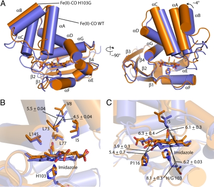
Heme nitric oxide/oxygen (H-NOX) proteins are found in eukaryotes where they are typically part of a larger protein such as soluble guanylate cyclase and in prokaryotes where they are often found in operons with a histidine kinase, suggesting that H-NOX proteins serve as sensors for NO and O(2) in signaling pathways. The Fe(II)-NO complex of the H-NOX protein from Shewanella oneidensis inhibits the autophosphorylation of the operon-associated histidine kinase, whereas the ligand-free H-NOX has no effect on the kinase. NMR spectroscopy was used to determine the structures of the Fe(II)-CO complex of the S. oneidensis H-NOX and the Fe(II)-CO complex of the H103G H-NOX mutant as a mimic of the ligand-free and kinase-inhibitory Fe(II)-NO H-NOX, respectively. The results provide a molecular glimpse into the ligand-induced conformational changes that may underlie kinase inhibition and the subsequent control of downstream signaling.


Conflict of interest statement

The authors declare no conflict of interest.

Figures

Fig. 1.
Fig. 1.
SO2144 H-NOX constructs for NMR studies. (A) Full-length SO2144 H-NOX is 181 residues as shown. The histidine kinase SO2145 is 311 residues including a dimerization and histidine phosphotransfer domain (DHp) and a catalytic and ATP-binding domain (CA). (B) WT SO2144 Fe(II)heme diatomic ligand complexes and the H103G SO2144 H-NOX mutant Fe(II)–CO complex. (C) Inhibition of the autophosphorylation reaction catalyzed by the SO2145 histidine kinase by Fe(II)–CO H103G.
Fig. 2.
Fig. 2.
Solution structures of the Fe(II)–CO WT and Fe(II)–CO H103G states. (A) Fe(II)–CO WT with the protein backbone and the side chain of proximal heme ligand H103 (blue); the heme cofactor with a distally bound CO ligand (yellow). (B) Fe(II)–CO H103G with the protein backbone (green) and the heme cofactor, the distal CO ligand, and proximal imidazole (purple). Residues are numbered according to their positions in the primary sequence.
Fig. 3.
Fig. 3.
Strictly conserved residues across all members of the H-NOX domain family. The residues in the heme-binding scaffold of the SO2144 H-NOX domain (H103, P116, Y132, S134, and R136) are conserved across the family, including those that induce heme nonplanarity. Residues that are strictly conserved in prokaryotic and eukaryotic H-NOX domains are colored in red.
Fig. 4.
Fig. 4.
Structure overlay with alignment of the proximal subdomain of Fe(II)–CO WT and Fe(II)–CO H103G. Fe(II)–CO WT is colored blue and Fe(II)-H103G is colored green. Backbone atoms of residues 100–110 and 120–178 in the Fe(II)–CO WT and Fe(II)–CO H103G structure ensembles were aligned.
Fig. 5.
Fig. 5.
Heme site structure. (A–C) (Upper) Fe(II)–CO WT. (Lower) Fe(II)–CO H103G. (A) The backbones of the Fe(II)–CO WT and Fe(II)–CO H103G structural ensembles are shown in blue and green and the heme cofactors are in yellow and purple, respectively. Residues 31–45 and 110–114 are omitted for clarity. Key residues that comprise the heme pocket are displayed in orange (I5, L77, H/G103, L115, P116, and L145). (B) Close-up view of the heme cofactor and the residues that comprise the heme site in the structural ensemble. (C) The heme site from the single structure in the NMR structural ensemble that is closest to the average conformation. (D) Heme deviations from planarity calculated using NSD (32). The average values of each mode across the ensembles are plotted in units of Å. (E) Box plots of selected NSD data.
Fig. 6.
Fig. 6.
Conformational differences between the Fe(II)–CO WT and Fe(II)–CO H103G states. (A) Overlay of the Fe(II)–CO WT and Fe(II)–CO H103G solution structures with the proximal subdomain, residues 100–110 (αF) and 120–178 (αG and β1–4), aligned. Fe(II)–CO WT is colored blue and Fe(II)–CO H103G is colored orange. (B) Key distance changes in the heme pocket distal to the heme. (C) Distance changes that underlie the conformational change after rupture of the axial histidine-iron bond. The single structures in each ensemble closest to the average are shown.

Similar articles

Cited by

References

    1. Derbyshire ER, Marletta MA. Biochemistry of soluble guanylate cyclase. Handb Exp Pharmacol. 2009:17–31. - PubMed
    1. Chan MK. Recent advances in heme-protein sensors. Curr Opin Chem Biol. 2001;5:216–222. - PubMed
    1. Boon EM, Huang SH, Marletta MA. A molecular basis for NO selectivity in soluble guanylate cyclase. Nat Chem Biol. 2005;1:53–59. - PubMed
    1. Huang SH, Rio DC, Marletta MA. Ligand binding and inhibition of an oxygen-sensitive soluble guanylate cyclase, Gyc-88E, from Drosophila. Biochemistry. 2007;46:15115–15122. - PubMed
    1. Iyer LM, Anantharaman V, Aravind L. Ancient conserved domains shared by animal soluble guanylyl cyclases and bacterial signaling proteins. BMC Genomics. 2003;4:5. - PMC - PubMed

Publication types

LinkOut - more resources