Review
. 2010 Jan;6(1):32-40.
doi: 10.1038/nrneph.2009.192. Epub 2009 Nov 17.

Bone disease after renal transplantation

Affiliations
Review

Bone disease after renal transplantation

Hartmut H Malluche et al. Nat Rev Nephrol. 2010 Jan.

Abstract

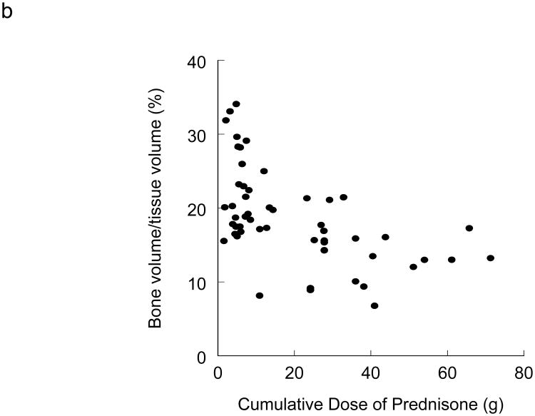
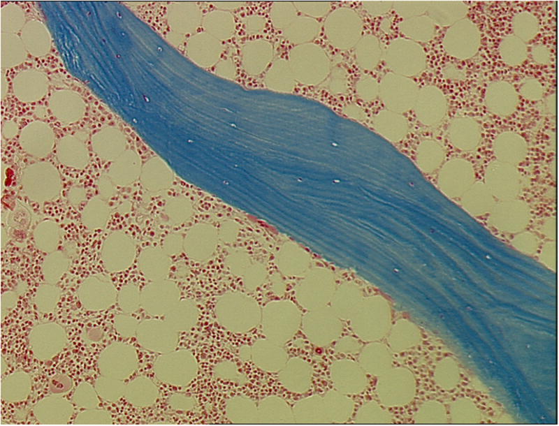
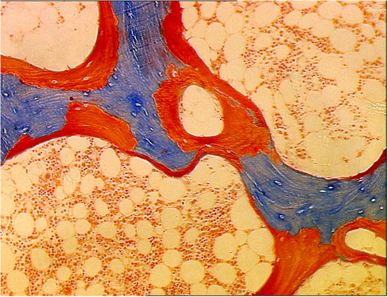
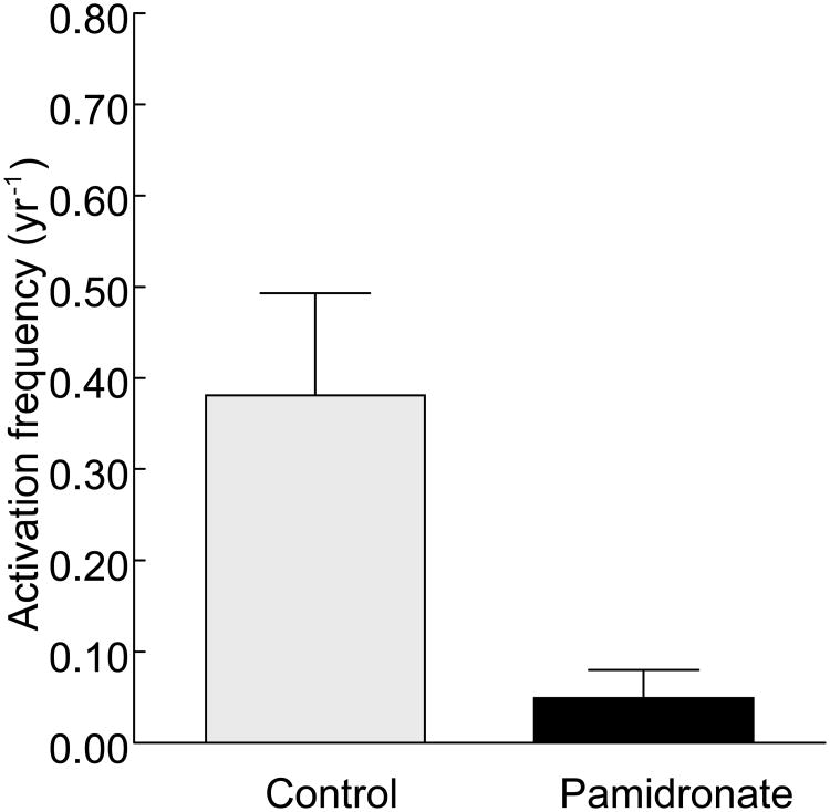
In light of greatly improved long-term patient and graft survival after renal transplantation, improving other clinical outcomes such as risk of fracture and cardiovascular disease is of paramount importance. After renal transplantation, a large percentage of patients lose bone. This loss of bone results from a combination of factors that include pre-existing renal osteodystrophy, immunosuppressive therapy, and the effects of chronically reduced renal function after transplantation. In addition to low bone volume, histological abnormalities include decreased bone turnover and defective mineralization. Low bone volume and low bone turnover were recently shown to be associated with cardiovascular calcifications, highlighting specific challenges for medical therapy and the need to prevent low bone turnover in the pretransplant patient. This Review discusses changes in bone histology and mineral metabolism that are associated with renal transplantation and the effects of these changes on clinical outcomes such as fractures and cardiovascular calcifications. Therapeutic modalities are evaluated based on our understanding of bone histology.


Figures

Figure 1
Figure 1
Glucocorticoids reduce bone turnover. a) Relationship between the cumulative dose of prednisone and activation frequency in 53 patients after renal transplantation. Coefficient of correlation = 0.43, P < 0.05. b) Relationship between cumulative dose of prednisone and BV/TV in 53 patients after renal transplantation. Coefficient of correlation = 0.54, P < 0.05. Abbreviation: BV/TV, bone volume fraction of total tissue volume. Permission obtained from American Society of Nephrology © Monier-Faugere, M. C. et al. High prevalence of low bone turnover and occurrence of osteomalacia after kidney transplantation. J. A. Soc. Nephrol. 11, 1093-1099 (2000).
Figure 1
Figure 1
Glucocorticoids reduce bone turnover. a) Relationship between the cumulative dose of prednisone and activation frequency in 53 patients after renal transplantation. Coefficient of correlation = 0.43, P < 0.05. b) Relationship between cumulative dose of prednisone and BV/TV in 53 patients after renal transplantation. Coefficient of correlation = 0.54, P < 0.05. Abbreviation: BV/TV, bone volume fraction of total tissue volume. Permission obtained from American Society of Nephrology © Monier-Faugere, M. C. et al. High prevalence of low bone turnover and occurrence of osteomalacia after kidney transplantation. J. A. Soc. Nephrol. 11, 1093-1099 (2000).
Figure 2
Figure 2
Low turnover bone disease (adynamic bone disease) in a 35-year-old white male renal transplant recipient. Bone biopsy was performed 24 months after kidney transplantation. No evidence of matrix apposition (osteoid) can be visualized. Only one osteoclast engaging in shallow resorption at the trabecular surface is evident (arrow). Mineralized, 4 μm thick bone section, stained with modified Masson-Goldner trichrome, original magnification ×100.
Figure 3
Figure 3
Low turnover osteomalacia in a 49-year-old white female renal transplant recipient. This bone biopsy was performed 14 months after kidney transplantation. High osteoid volume (arrows) secondary to increased osteoid surface and thick osteoid seams is evident. No osteoblasts or osteoclasts are present. Mineralized, 4 μm thick bone section, stained with modified Masson-Goldner trichrome, original magnification ×100.
Figure 4
Figure 4
Administration of pamidronate reduces bone turnover after renal transplantation. Activation frequency was assessed by histomorphometry in patients 6 months after renal transplantation. Patients in both the control group and the pamidronate group received low-dose calcitriol and calcium carbonate. Patients in the pamidronate group received 60 mg intravenous pamidronate immediately after renal transplantation followed by 30 mg at months 1, 2 and 6.

References

    1. Nishikawa K, Terasaki PI. Annual trends and triple therapy--1991-2000. Clin Transpl. 2001:247–69. - PubMed
    1. Marcen R, Teruel JL. Patient outcomes after kidney allograft loss. Transplant Rev (Orlando) 2008;22:62–72. - PubMed
    1. Malluche HH, Faugere MC. Atlas of Mineralized Bone Histology. Karger; New York: 1986.
    1. Hruska KA, Teitelbaum SL. Renal osteodystrophy. N Engl J Med. 1995;333:166–74. - PubMed
    1. Wang M, et al. Relationship between intact 1-84 parathyroid hormone and bone histomorphometric parameters in dialysis patients without aluminum toxicity. Am J Kidney Dis. 1995;26:836–44. - PubMed

Publication types

MeSH terms