

Induction of antigen-specific immune tolerance by TGF-beta-induced CD4+Foxp3+ regulatory T cells
- PMID: 19918314
- PMCID: PMC2770184
Induction of antigen-specific immune tolerance by TGF-beta-induced CD4+Foxp3+ regulatory T cells
Abstract
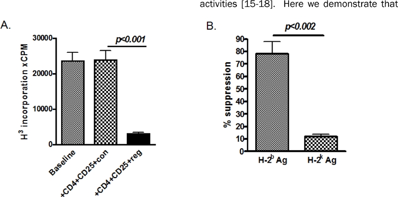
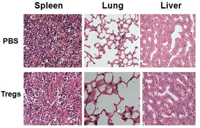
Like natural CD4(+)CD25(+) Treg cells, TGF-beta-induced Treg cells also prevent allograft rejection in MHC-mismatched organ transplantation models. In analyzing this effect with greater detail, we determined that injection of TGF-beta-induced, alloactivated CD4(+)CD25(+) cells induces antigen-specific immune tolerance in vivo. Increased CD4(+)CD25(+) cells in recipients contribute to this immune tolerance. In addition, adoptive transfer of TGF-beta-induced CD4(+)CD25(+) cells did not result in significant toxic and side effects in recipients. These results indicate that TGF-beta-induced, alloactivated CD4(+)CD25(+) cells may provide a safe and effective approach to protect MHC-mismatched organ grafts from rejection in a clinical setting.
Keywords: Foxp3; Immunoregulation; TGF-β; regulatory T cells; transplant tolerance.
Figures


Similar articles
-
Transfer of regulatory T cells generated ex vivo modifies graft rejection through induction of tolerogenic CD4+CD25+ cells in the recipient.Int Immunol. 2006 Feb;18(2):279-89. doi: 10.1093/intimm/dxh368. Epub 2006 Jan 13. Int Immunol. 2006. PMID: 16415106
-
PD-L1 signal on liver dendritic cells is critical for Foxp3(+)CD4(+)CD25(+) Treg and liver tolerance induction in mice.Transplant Proc. 2013 Jun;45(5):1853-5. doi: 10.1016/j.transproceed.2013.03.015. Transplant Proc. 2013. PMID: 23769057
-
New insights into mechanisms of spontaneous liver transplant tolerance: the role of Foxp3-expressing CD25+CD4+ regulatory T cells.Am J Transplant. 2008 Aug;8(8):1639-51. doi: 10.1111/j.1600-6143.2008.02300.x. Epub 2008 Jun 12. Am J Transplant. 2008. PMID: 18557727
-
Alloantigen specific T regulatory cells in transplant tolerance.Int Immunopharmacol. 2009 May;9(5):570-4. doi: 10.1016/j.intimp.2009.01.016. Epub 2009 Jan 29. Int Immunopharmacol. 2009. PMID: 19539571 Review.
-
CD4+CD25+ T regulatory cells in renal transplantation.Front Immunol. 2022 Nov 8;13:1017683. doi: 10.3389/fimmu.2022.1017683. eCollection 2022. Front Immunol. 2022. PMID: 36426347 Free PMC article. Review.
Cited by
-
Thioredoxin priming prolongs lung allograft survival by promoting immune tolerance.PLoS One. 2015 May 1;10(5):e0124705. doi: 10.1371/journal.pone.0124705. eCollection 2015. PLoS One. 2015. PMID: 25933390 Free PMC article.
-
Increased prevalence of regulatory T cells in the lung cancer microenvironment: a role of thymic stromal lymphopoietin.Cancer Immunol Immunother. 2011 Nov;60(11):1587-96. doi: 10.1007/s00262-011-1059-6. Epub 2011 Jun 17. Cancer Immunol Immunother. 2011. PMID: 21681373 Free PMC article.
-
Characterization of Schistosoma japonicum CP1412 protein as a novel member of the ribonuclease T2 molecule family with immune regulatory function.Parasit Vectors. 2017 Feb 17;10(1):89. doi: 10.1186/s13071-016-1962-y. Parasit Vectors. 2017. PMID: 28212670 Free PMC article.
-
Immunomodulatory Function of Vitamin D and Its Role in Autoimmune Thyroid Disease.Front Immunol. 2021 Feb 19;12:574967. doi: 10.3389/fimmu.2021.574967. eCollection 2021. Front Immunol. 2021. PMID: 33679732 Free PMC article. Review.
-
Murine regulatory T cells utilize granzyme B to promote tumor metastasis.Cancer Immunol Immunother. 2023 Sep;72(9):2927-2937. doi: 10.1007/s00262-023-03410-w. Epub 2023 Feb 24. Cancer Immunol Immunother. 2023. PMID: 36826509 Free PMC article.
References
-
- Sakaguchi S, Yamaguchi T, Nomura T, Ono M. Regulatory T cells and immune tolerance. Cell. 2008;133:775–787. - PubMed
-
- Kang SM, Tang Q, Bluestone JA. CD4+CD25+ regulatory T cells in transplantation: progress, challenges and prospects. Am J Transplant. 2007;7:1457–1463. - PubMed
-
- Dijke IE, Korevaar SS, Caliskan K, Balk AH, Maat AP, Weimar W, Baan CC. Inadequate immune regulatory function of CD4+CD25 bright+FoxP3+ T cells in heart transplant patients who experience acute cellular rejection. Transplantation. 2009;87:1191–1200. - PubMed
-
- Trenado A, Sudres M, Tang Q, Maury S, Charlotte F, Grégoire S, Bonyhadi M, Klatzmann D, Salomon BL, Cohen JL. Ex vivo-expanded CD4+CD25+ immunoregulatory T cells prevent graft-versus-host-disease by inhibiting activation/differentiation of pathogenic T cells. J Immunol. 2006;176:1266–1273. - PubMed
LinkOut - more resources
Full Text Sources
Research Materials