Dramatic co-activation of WWOX/WOX1 with CREB and NF-kappaB in delayed loss of small dorsal root ganglion neurons upon sciatic nerve transection in rats
- PMID: 19918364
- PMCID: PMC2771921
- DOI: 10.1371/journal.pone.0007820
Dramatic co-activation of WWOX/WOX1 with CREB and NF-kappaB in delayed loss of small dorsal root ganglion neurons upon sciatic nerve transection in rats
Abstract
Background: Tumor suppressor WOX1 (also named WWOX or FOR) is known to participate in neuronal apoptosis in vivo. Here, we investigated the functional role of WOX1 and transcription factors in the delayed loss of axotomized neurons in dorsal root ganglia (DRG) in rats.
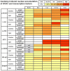
Methodology/principal findings: Sciatic nerve transection in rats rapidly induced JNK1 activation and upregulation of mRNA and protein expression of WOX1 in the injured DRG neurons in 30 min. Accumulation of p-WOX1, p-JNK1, p-CREB, p-c-Jun, NF-kappaB and ATF3 in the nuclei of injured neurons took place within hours or the first week of injury. At the second month, dramatic nuclear accumulation of WOX1 with CREB (>65% neurons) and NF-kappaB (40-65%) occurred essentially in small DRG neurons, followed by apoptosis at later months. WOX1 physically interacted with CREB most strongly in the nuclei as determined by FRET analysis. Immunoelectron microscopy revealed the complex formation of p-WOX1 with p-CREB and p-c-Jun in vivo. WOX1 blocked the prosurvival CREB-, CRE-, and AP-1-mediated promoter activation in vitro. In contrast, WOX1 enhanced promoter activation governed by c-Jun, Elk-1 and NF-kappaB. WOX1 directly activated NF-kappaB-regulated promoter via its WW domains. Smad4 and p53 were not involved in the delayed loss of small DRG neurons.
Conclusions/significance: Rapid activation of JNK1 and WOX1 during the acute phase of injury is critical in determining neuronal survival or death, as both proteins functionally antagonize. In the chronic phase, concurrent activation of WOX1, CREB, and NF-kappaB occurs in small neurons just prior to apoptosis. Likely in vivo interactions are: 1) WOX1 inhibits the neuroprotective CREB, which leads to eventual neuronal death, and 2) WOX1 enhances NF-kappaB promoter activation (which turns to be proapoptotic). Evidently, WOX1 is the potential target for drug intervention in mitigating symptoms associated with neuronal injury.
Conflict of interest statement
Figures


Similar articles
-
Zfra affects TNF-mediated cell death by interacting with death domain protein TRADD and negatively regulates the activation of NF-kappaB, JNK1, p53 and WOX1 during stress response.BMC Mol Biol. 2007 Jun 13;8:50. doi: 10.1186/1471-2199-8-50. BMC Mol Biol. 2007. PMID: 17567906 Free PMC article.
-
MPP+-induced neuronal death in rats involves tyrosine 33 phosphorylation of WW domain-containing oxidoreductase WOX1.Eur J Neurosci. 2008 Apr;27(7):1634-46. doi: 10.1111/j.1460-9568.2008.06139.x. Epub 2008 Mar 26. Eur J Neurosci. 2008. PMID: 18371080
-
The transcription factors c-JUN, JUN D and CREB, but not FOS and KROX-24, are differentially regulated in axotomized neurons following transection of rat sciatic nerve.Brain Res Mol Brain Res. 1992 Jul;14(3):155-65. doi: 10.1016/0169-328x(92)90170-g. Brain Res Mol Brain Res. 1992. PMID: 1331648
-
Role of WWOX and NF-κB in lung cancer progression.Transl Respir Med. 2013 Dec;1(1):15. doi: 10.1186/2213-0802-1-15. Epub 2013 Nov 14. Transl Respir Med. 2013. PMID: 27234396 Free PMC article. Review.
-
Role of WWOX/WOX1 in Alzheimer's disease pathology and in cell death signaling.Front Biosci (Schol Ed). 2013 Jan 1;5(1):72-85. doi: 10.2741/s358. Front Biosci (Schol Ed). 2013. PMID: 23277037 Review.
Cited by
-
HGF and TGFβ1 differently influenced Wwox regulatory function on Twist program for mesenchymal-epithelial transition in bone metastatic versus parental breast carcinoma cells.Mol Cancer. 2015 Jun 4;14:112. doi: 10.1186/s12943-015-0389-y. Mol Cancer. 2015. Retraction in: Mol Cancer. 2020 Aug 15;19(1):126. doi: 10.1186/s12943-020-01242-1. PMID: 26041563 Free PMC article. Retracted.
-
WWOX dysfunction induces sequential aggregation of TRAPPC6AΔ, TIAF1, tau and amyloid β, and causes apoptosis.Cell Death Discov. 2015 Aug 3;1:15003. doi: 10.1038/cddiscovery.2015.3. eCollection 2015. Cell Death Discov. 2015. PMID: 27551439 Free PMC article.
-
Systematic analysis of expression signatures of neuronal subpopulations in the VTA.Mol Brain. 2019 Dec 11;12(1):110. doi: 10.1186/s13041-019-0530-8. Mol Brain. 2019. PMID: 31829254 Free PMC article.
-
Modeling WWOX Loss of Function in vivo: What Have We Learned?Front Oncol. 2018 Oct 10;8:420. doi: 10.3389/fonc.2018.00420. eCollection 2018. Front Oncol. 2018. PMID: 30370248 Free PMC article. Review.
-
WWOX Controls Cell Survival, Immune Response and Disease Progression by pY33 to pS14 Transition to Alternate Signaling Partners.Cells. 2022 Jul 7;11(14):2137. doi: 10.3390/cells11142137. Cells. 2022. PMID: 35883580 Free PMC article. Review.
References
-
- Sze CI, Su M, Pugazhenthi S, Jambal P, Hsu LJ, et al. Downregulation of WW domain-containing oxidoreductase induces Tau phosphorylation in vitro: A potential role in Alzheimer's disease. J Biol Chem. 2004;279:30498–30506. - PubMed
-
- Chen ST, Chuang JI, Cheng CL, Hsu LJ, Chang NS. Light-induced retinal damage involves tyrosine 33 phosphorylation, mitochondrial and nuclear translation of WW domain-containing oxidoreductase in vivo. Neuroscience. 2005;130:397–407. - PubMed
-
- Lo CP, Hsu LJ, Li MY, Hsu SY, Chuang JI, et al. MPP+-Induced neuronal death in rats involves tyrosine 33 phosphorylation of WW domain-containing oxidoreductase WOX1. Eur J Neurosci. 2008;27:1634–1646. - PubMed
-
- Bednarek AK, Laflin KJ, Daniel RL, Liao Q, Hawkins KA, et al. WWOX, a novel WW domain-containing protein mapping to human chromosome 16q23.3-24.1, a region frequently affected in breast cancer. Cancer Res. 2000;60:2140–2145. - PubMed
-
- Ried K, Finnis M, Hobson L, Mangelsdorf M, Dayan S, et al. Common chromosomal fragile site FRA16D sequence: identification of the FOR gene spanning FRA16D and homozygous deletions and translocation breakpoints in cancer cells. Hum Mol Genet. 2000;9:1651–1663. - PubMed
Publication types
MeSH terms
Substances
Grants and funding
LinkOut - more resources
Full Text Sources
Research Materials
Miscellaneous

