Case Reports
. 2009 Aug 25:2:7143.
doi: 10.4076/1757-1626-2-7143.

Brucella liver abscess; imaging approach, differential diagnosis, and therapeutic management: a case report

Affiliations
Case Reports

Brucella liver abscess; imaging approach, differential diagnosis, and therapeutic management: a case report

Danai Chourmouzi et al. Cases J. .

Abstract

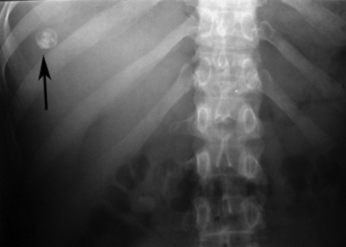
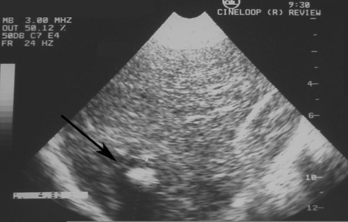
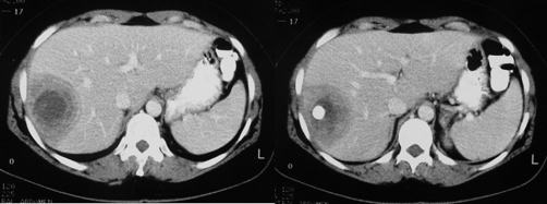
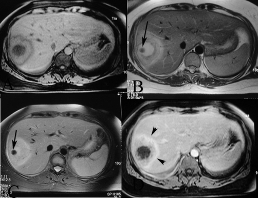
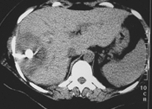
We report a new case of a brucellar liver abscess (brucelloma) in a young woman without previous remote brucellosis who presented with pronounced systemic and mild local symptoms. Brucelloma is the result of calcification of a granoulomatous reaction induced by persistent Brucella in macrophages. It represents a rare manifestation that follows previously undetected brucellosis. We describe the findings in plain radiograph, ultrasound, computed tomography, and magnetic resonance images. Together with the positive serology, imaging yielded important elements supporting the diagnosis. Modern radiological techniques also contributed to the final therapeutic management, preventing unnecessary laparotomy. Sequencing confirmed the definite diagnosis of Brucella melitensis as the causative factor.


Figures

Figure 1.
Figure 1.
Plain film of the right hypochondrium shows a well defined, rounded calcification with an inner snowflake pattern.
Figure 2.
Figure 2.
Ultrasound of the liver reveals a central hyperechoic lesion surrounded by a hypoechoic halo with ill-defined borders.
Figure 3.
Figure 3.
Contrast-enhanced CT scan reveals a hypoattenuating lesion in the right hepatic lobe with peripheral enhanced thickened wall. A central dense calcium deposit is also observed.
Figure 4.
Figure 4.
Axial MR images reveal (a) a central hypo-intense lesion on vibe, and hyperintensity on (b) T2-weighted and (c) STIR, with enchanced halo on gadolinium-enhanced images. (d) Periabscess liver tissue is hypointense on T1-weighted and mildly hyperintense on T2-weighted images, and reveals increased enhancement on post-gadolinium images (arrowheads). Note the dense calcium deposit as a low signal intensity lesion (arrows).
Figure 5.
Figure 5.
CT scan during percutaneous drainage under CT guidance reveals that the drainage catheter is in good position within the abscess cavity.

References

    1. Akritidis N, Tzivras M, Delladetsima I, Stefanaki S, Moutsopoulos HM, Pappas G. The liver in brucellosis. Clin Gastroenterol Hepatol. 2007;5(9):1109–1112. doi: 10.1016/j.cgh.2006.08.010. - DOI - PubMed
    1. Sadia Pérez D, Cea-Calvo L, Aguado García JM, Ruiz Ilundain G, López Martín A, González Gómez C. Brucella hepatic abscess. Report of a case and review of the literature. Rev Clin Esp. 2001;201(6):322–326. - PubMed
    1. Rovery C, Rolain JM, Raoult D, Brouqui P. Shell vial culture as a tool for isolation of Brucella melitensis in chronic hepatic abscess. Clin Microbiol. 2003;41(9):4460–4461. doi: 10.1128/JCM.41.9.4460-4461.2003. - DOI - PMC - PubMed
    1. Sisteron O, Souci J, Chevallier P, Cua E, Bruneton JN. Hepatic abscess caused by Brucella US, CT and MRI findings: case report and review of the literature. Clin Imaging. 2002;26(6):414–417. doi: 10.1016/S0899-7071(02)00507-7. - DOI - PubMed
    1. Foulongne V, Bourg G, Cazevieille C, Michaux-Charachon S, O'Callaghan D. Identification of Brucella suis genes affecting intracellular survival in an in vitro human macrophage infection model by signature-tagged transposon mutagenesis. Infect Immun. 2000;68(3):1297–1303. doi: 10.1128/IAI.68.3.1297-1303.2000. - DOI - PMC - PubMed

Publication types

LinkOut - more resources