Case Reports
. 2009 Sep 15:2:6784.
doi: 10.4076/1757-1626-2-6784.

Conservative management for an esophageal perforation in a patient presented with delayed diagnosis: a case report review of the literature

Affiliations
Case Reports

Conservative management for an esophageal perforation in a patient presented with delayed diagnosis: a case report review of the literature

Konstantinos Tsalis et al. Cases J. .

Abstract

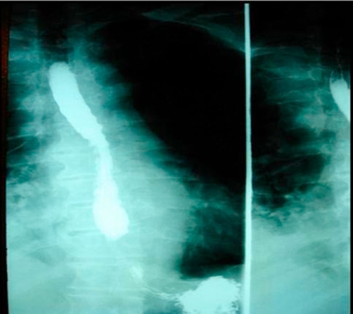
Esophageal perforation is a serious condition with a high mortality rate. Successful therapy depends on the size of the rupture; the time elapsed between rupture and diagnosis, and the underlying health of the patient. Common causes of esophageal perforation include medical instrumentation, foreign-body ingestion, and trauma. A case of esophageal perforation due to fish bone ingestion in a 67-year-old male is described here, with a review of the pertinent literature. The patient presented with chest pain, fever and right-sided pleural effusion. Initial evaluation was nondiagnostic. The water-soluble contrast swallow test showed no evidence of leakage. Computed tomography scan demonstrated a pneumomediastinum, and right-sided hydropneumothorax. The patient was successfully treated using conservative measures.


Figures

Figure 1.
Figure 1.
Chest X-ray demonstrated right pleural effusion, but no radio-opacity was detected and there was no evidence of pneumomediastinum or subcutaneous emphysema.
Figure 2.
Figure 2.
A hypaque swallow study failed to demonstrate extravasation of contrast medium.
Figure 3.
Figure 3.
Erect chest X-ray a few hours later demonstrated contrast medium extravasation accompanied with large pleural effusion.
Figure 4.
Figure 4.
Subsequent CT scan demonstrated right sided pneumothorax, extended right sided pleural effusion and a small amount of air in the mediastinum.
Figure 5.
Figure 5.
A confirmative esophagogastroduodenoscopy revealed a small distal esophageal perforation.
Figure 6.
Figure 6.
A covered self-expanding metallic stent was inserted endoscopically, across the tear site to prevent ongoing local infection.
Figure 7.
Figure 7.
Follow up CT scan at 3 months.

References

    1. Kanowitz A, Markovchick V. Oesophageal and diaphragmatic trauma. In: Rosen P, editor. Emergency medicine: concepts and clinical practice. 4. St Louis: Mosby; 1998. pp. 546–548.
    1. Nandi P, Ong GB. Foreign body in the oesophagus: review of 2394 cases. Br J Surg. 1978;65:5–9. doi: 10.1002/bjs.1800650103. - DOI - PubMed
    1. Persaud RA, Sudhakaran N, Ong CC, Bowdler DA, Dykes E. Extraluminal migration of a coin in the oesophagus of a child misdiagnosed as asthma. Emerg Med J. 2001;18:312–313. doi: 10.1136/emj.18.4.312. - DOI - PMC - PubMed
    1. Dahiya M, Denton JS. Esophagoaortic perforation by foreign body (coin) causing sudden death in a 3-year-old child. Am J Forensic Med Pathol. 1999;20:184–188. doi: 10.1097/00000433-199906000-00016. - DOI - PubMed
    1. Theodoropoulou R, Roussomoustakaki M, Michalodimitrakis MN, Kanaki C, Kouroumalis EA. Fatal hepatic abscess caused by a fish bone. Lancet. 2002;359:977. doi: 10.1016/S0140-6736(02)07999-0. - DOI - PubMed

Publication types

LinkOut - more resources