. 2010 May;28(5):687-96.
doi: 10.1002/jor.21033.

Recombinant human bone morphogenetic protein-7 enhances fracture healing in an ischemic environment

Affiliations

Recombinant human bone morphogenetic protein-7 enhances fracture healing in an ischemic environment

Chuanyong Lu et al. J Orthop Res. 2010 May.

Abstract

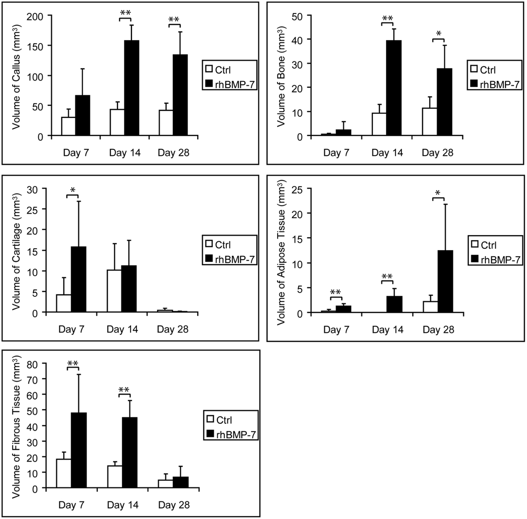
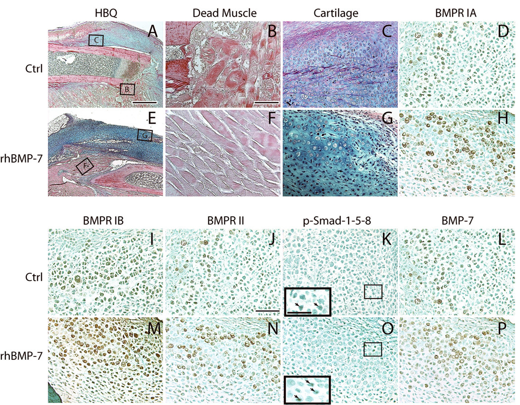
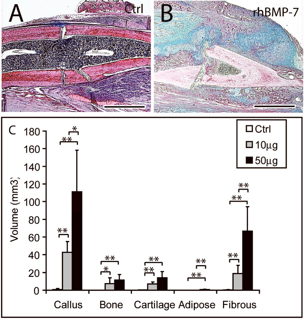
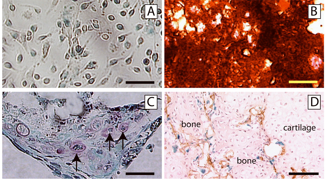
Ischemia predisposes orthopedic trauma patients to delayed fracture healing or nonunion. The goal of this study was to test the ability of bone morphogenetic protein 7 (BMP7) to stimulate fracture repair in an ischemic environment. Ischemic fractures were generated in male adult mice by resecting the femoral artery prior to the creation of a nonstabilized tibia fracture. Recombinant human BMP7 (rhBMP7, 50 microg) was injected into the fracture site immediately after surgery. At 7 days after injury, more tissue vascularization was observed in rhBMP7 treated fractures. Histomorphometric analyses revealed that rhBMP7 induced more cartilage at day 7, more callus and bone at days 14 and 28, and more adipose tissue and fibrous tissue at days 7, 14, and 28 compared to controls (n=5/group/time). At day 28, all fractures treated with rhBMP7 (50 microg, n=5) healed, whereas only three of five control fractures exhibited slight bony bridging. In addition, we found that rhBMP7 (both 10 and 50 microg) significantly increased the amount of cartilage compared to controls in stabilized fractures, confirming its chondrogenic effect. Lastly, using bone marrow transplantation, we determined that no donor-derived osteocytes or chondrocytes were present in rhBMP7-treated fractures, suggesting rhBMP7 did not recruit mesenchymal stem cells from the bone marrow to the fracture site. In conclusion, our results indicate that rhBMP7 is a promising treatment for fractures with severely disrupted blood supply.


Figures

Fig. 1
Fig. 1. Histomorphometric analyses of fracture healing in rhBMP7 treated ischemic fractures
Animals with tibia fracture and femoral artery resection were treated with 50µg of rhBMP7. * p<0.05, ** p<0.01.
Fig. 2
Fig. 2. Increased activation of Bmp signaling pathway after 50µg of rhBMP7 treatment: 7 days after fracture and femoral artery resection
(A-D, I-L) Representative micrographs of control fractures. (A) A fracture callus stained by Hall’s and Brunt’s Quadruple stain method (HBQ). Cartilage appears blue. (B) Necrotic myofibers at the fracture site. (C) A cartilage island formed in the callus. (D, I-L) Bmp receptors IA (BMPR IA), IB, II, phosphorylated Smad-1-5-8, and Bmp7 are detected in chondrocytes. Insert in (K) is a high magnification of the box area, showing p-Smad-1-5-8 positive cells (arrows). (E-H, M-P) Representative micrographs of ischemic fractures treated with rhBMP7. (E) A fracture callus of rhBMP7 treated fracture. (F) Necrotic myofibers at the fracture site. (G) A cartilage island. (H, M-P) rhBMP7 treated fractures have stronger immunostaining for Bmp receptors IA, IB, II, phosphorylated Smad-1-5-8, and Bmp7 in chondrocytes compared to the control. Insert in (O) is a high magnification of the box area, showing p-Smad-1-5-8 positive cells (arrows). Scale bars: (A, E) = 1mm, (B-D, F-H, I-L, M-P) = 100µm, inserts in (K and O) = 50 µm.
Fig. 3
Fig. 3. Histology of rhBMP7 treated ischemic fractures at 14 days after injury: HBQ staining
(A) A representative fracture callus of control animals. (B) New bone (red) in control fractures is well organized. (C) Fibrous tissue is present between dead muscle fibers (m). (D) A representative fracture callus of rhBMP7 treated animals. (E) rhBMP7-induced new bone has a different morphology compared to trabeculae in control fractures (B). (F) Cartilage islands (blue) are observed between dead muscle fibers (m). Scale bars: (A and D) = 2mm, (B, E) = 200µm, (C, F) = 100µm.
Fig. 4
Fig. 4. Histology of rhBMP7 treated ischemic fractures at 28 days after injury: HBQ staining
(A) In the controls, fibrous tissue (outlined areas with solid line) and cartilage (blue) are present in callus. (B) rhBMP7 treated fractures have large bony calluses with minimal amount of fibrous tissue and cartilage. Area outlined with dashed line is fracture callus. Scale bar = 2mm.
Fig. 5
Fig. 5. Stabilized fractures treated with rhBMP7
Fracture healing was analyzed at 10 days after injury. (A) A representative callus of the controls showing minimal bone formation at fracture site. (B) Fifty micrograms of rhBMP7 induces a large amount of bone and cartilage formation. (C) Histomorphometric analyses. Scale bar = 1mm. * p<0.05, ** p<0.01.
Fig. 6
Fig. 6. Osteogenic/chondrogenic progenitor cells are not recruited from bone marrow in rhBMP7 treated fractures
(A) Bone marrow cells harvested from chimeric mice formed colonies. A majority of cells in the colony are positive for X-gal staining (blue staining). (B) These colony forming cells differentiated into osteogenic cells, producing mineralized matrix which is stained red by Alizarin Red solution. (C) These colony forming cells were also capable of differentiating into chondrocytes (arrows, Safranin O/Fast Green staining). (D) Ischemic tibia fracture was created in chimeric mice and treated with 50µg of rhBMP7. At 14 days after injury, blood vessels (brown, PECAM immunohistochemistry) invaded a cartilage island (cartilage), with new bone forming (bone). Chondrocytes in the cartilage, osteocytes in the new bone, and endothelial cells (brown staining) are negative for X-gal staining. Some X-gal positive cells (blue) are present at the endochondral front. Scale bars: A, C = 50µm, B = 200µm, D = 100µm.

References

    1. Dickson KF, Katzman S, Paiement G. The importance of the blood supply in the healing of tibial fractures. Contemp Orthop. 1995;30:489–493. - PubMed
    1. Einhorn TA. Enhancement of fracture-healing. J Bone Joint Surg Am. 1995;77:940–956. - PubMed
    1. Lu C, Miclau T, Hu D, Marcucio RS. Ischemia leads to delayed union during fracture healing: a mouse model. J Orthop Res. 2007;25:51–61. - PMC - PubMed
    1. Lu C, Rollins M, Hou H, et al. Tibial fracture decreases oxygen levels at the site of injury. Iowa Orthop J. 2008;28:14–21. - PMC - PubMed
    1. Hak DJ, Makino T, Niikura T, et al. Recombinant human BMP-7 effectively prevents nonunion in both young and old rats. J Orthop Res. 2006;24:11–20. - PubMed

Publication types

MeSH terms

Substances