. 2010 Jan;239(1):327-37.
doi: 10.1002/dvdy.22156.

WNT5A mutations in patients with autosomal dominant Robinow syndrome

Affiliations

WNT5A mutations in patients with autosomal dominant Robinow syndrome

Anthony D Person et al. Dev Dyn. 2010 Jan.

Abstract

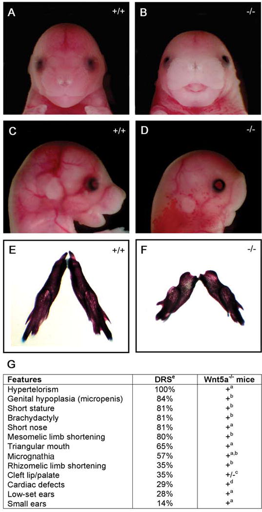
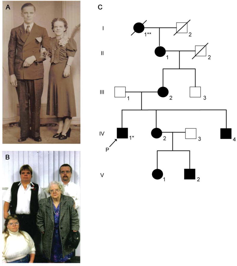
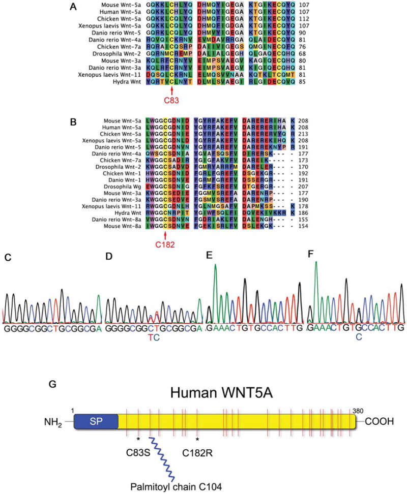
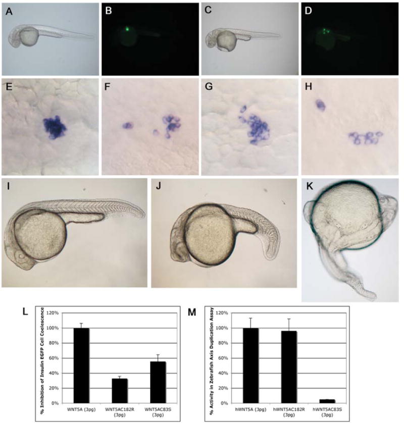
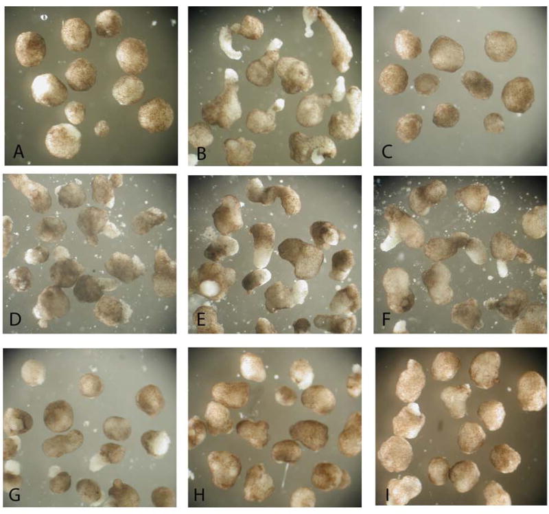
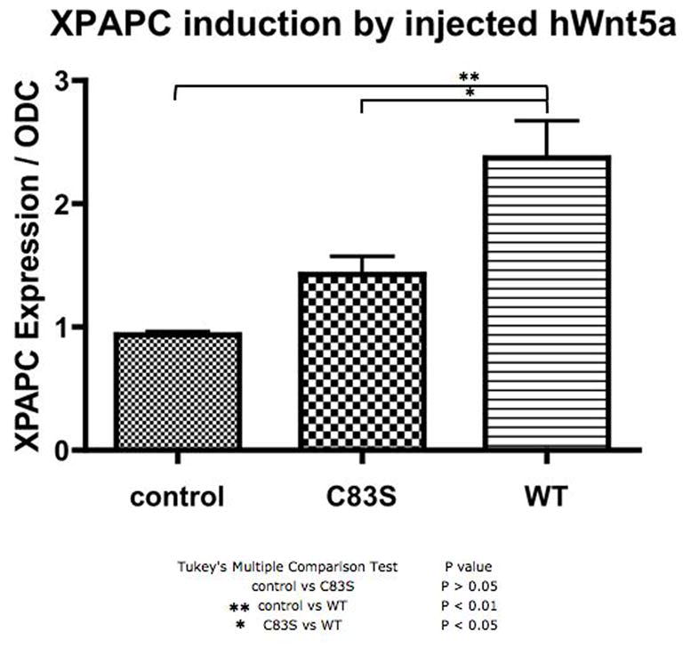
Robinow syndrome is a skeletal dysplasia with both autosomal dominant and autosomal recessive inheritance patterns. It is characterized by short stature, limb shortening, genital hypoplasia, and craniofacial abnormalities. The etiology of dominant Robinow syndrome is unknown; however, the phenotypically more severe autosomal recessive form of Robinow syndrome has been associated with mutations in the orphan tyrosine kinase receptor, ROR2, which has recently been identified as a putative WNT5A receptor. Here, we show that two different missense mutations in WNT5A, which result in amino acid substitutions of highly conserved cysteines, are associated with autosomal dominant Robinow syndrome. One mutation has been found in all living affected members of the original family described by Meinhard Robinow and another in a second unrelated patient. These missense mutations result in decreased WNT5A activity in functional assays of zebrafish and Xenopus development. This work suggests that a WNT5A/ROR2 signal transduction pathway is important in human craniofacial and skeletal development and that proper formation and growth of these structures is sensitive to variations in WNT5A function.


Conflict of interest statement

Competing Interest Statement

The authors declare that they have no competing financial interests.

Figures

Figure 1
Figure 1. Wnt5a−/− mice have anatomical defects phenotypically similar to patients with autosomal dominant Robinow syndrome
Wnt5a−/− mouse embryos at E16.5 (B) have widely spaced eyes (hypertelorism) and a triangular mouth compared to controls (A). Lateral views (C, D) reveal flattening of the facial profile, micrognathia, and small, low set ears in the Wnt5a−/− embryos (D) compared to control littermates (C). Skeletal preparations at birth show mandibular hypoplasia in Wnt5a mutants (F) compared to controls (E). G) Percentage of reported findings in dominant Robinow syndrome patients compared to phenotypes in Wnt5a−/− mice. aData presented in this figure, b (Yamaguchi et al., 1999) c(Yang et al., 2003) d(Schleiffarth et al., 2007). ePercentages of dominant Robinow syndrome (DRS) patients with each abnormality was adapted from ((Mazzeu et al., 2007b).
Figure 2
Figure 2. Index Family – Robinow syndrome
A) Photograph of an unaffected male (II-2) and his wife (II-1) with dominant Robinow syndrome. Features evident in the affected female include short stature, hypertelorism, and mesomelic limb shortening. B) Proband (Robinow et al., 1969), siblings and grandmother, who is the female pictured in panel A. Photograph in panel B from left to right, top row: male IV-4, male IV-1 (*proband); bottom row: female IV-2, and female II-1. Robinow syndrome features (described above) are evident in all four individuals. Additional images of all four individuals have been reported previously (Robinow et al., 1969). C) Pedigree of the index family with dominant Robinow syndrome originally described in (Robinow et al., 1969). An additional generation (V) is shown in this pedigree. ** Original pedigree reported male I-2 and not female I-1 had dominant Robinow syndrome (Robinow et al., 1969). We confirmed female I-1, and not male I-2, had dominant Robinow syndrome by consulting the family.
Figure 3
Figure 3. Dominant Robinow syndrome is associated with C83S or C182R WNT5A mutations
A, B) Sequence alignments show the cysteines (C83 and C182, red arrows) are conserved from human to hydra. C) Sequence data of a WNT5A exon 4 PCR product showing the wild type sequence of base pairs 537–552. D) Exon 4 PCR sequence data of a patient IV-1 showing a missense heterozygous mutation 544-545CT→TC (C182R). E) Control PCR sequence data of WNT5A bases 240–255. F) Sequence of an individual with autosomal dominant Robinow syndrome with a missense heterozygous mutation 248G→C (C83S). G) Schematic of the human WNT5A protein with a signal peptide (SP), 24 cysteines shown in red with the C83S and C182R mutation sites * and the palmitoylation site on C104 (Kurayoshi et al., 2007).
Figure 4
Figure 4. Cysteine mutations in WNT5A reduce the ability of this protein to activate non-canonical Wnt signaling in zebrafish embryos
Uninjected brightfield (A) and fluorescent images of insulin-eGFP transgenic zebrafish embryos (B) at 30hpf showing insulin-positive cells have coalesced to form an islet. Insulin-eGFP positive cells in embryos injected with WNT5A mRNA (C, D) do not coalesce properly, providing an in vivo assay for non-canonical Wnt5a activity. In situ hybridization showing insulin-expressing cells coalescing to form the zebrafish islet at 30hpf (E). Injections with WNT5A mRNA (F), WNT5AC182R mRNA (G, H) or WNT5AC83R mRNA (not shown) show scattered insulin expressing cells at 30 hpf, but the efficacy of this effect is reduced in WNT5AC182R (32%±3% n=93 P=0.00005) and WNT5AC83R (55%±9% n=152 P=0.002) mRNA injected embryos compared to WNT5A mRNA injected embryos (100%±6% n=260) (L). I) Uninjected zebrafish embryo at 26hpf. J) Zebrafish embryo at 26hpf injected with dnWNT5A mRNA (25pg) showing a bent tail phenotype similar to pipetail (Wnt5) mutants. Injections of WNT5A mRNA (K) or WNT5AC182R mRNA (not shown) into zebrafish embryos at the one-cell stage results in duplications of the embryonic axis, providing an overexpression-based in vivo activity assay for Wnt signaling competency by the WNT5A sequence variants. Normalizing the axis duplicating activity of WNT5A mRNA injected embryos to 100%±13% n=196, we noted indistinguishable axis duplicating activity 96%±16% n=65 in WNT5AC182 mRNA injected embryos (M). Injections of 3pg of WNT5AC83S mRNA resulted in a significantly different (P=0.001), low percentage of axis duplication activity 3%±0% n=102 compared to the full duplicating activity of WNT5A mRNA at the same dose (3pg) (M). In contrast, dnWNT5A injections did not duplicate the embryonic axis (J).
Figure 5
Figure 5. WNT5AC182R and WNT5AC182S have reduced ability to block activin-mediated cell movements during Xenopus gastrulation
Control (uninjected) blastula stage animal caps round up in normal culture conditions (A) and elongate when cultured in activin protein (B). Embryos injected with 200 pg of WNT5AC182R mRNA into each blastomere at a two cell stage form normal appearing animal caps under control conditions (C). Moderate dose (30 pg) WNT5A mRNA (D) and High dose (200 pg WNT5A mRNA) (G) block activin-induced animal cap elongation. In contrast, moderate dose (30 pg) WNT5AC182 mRNA (E) and WNT5AC83S mRNA (F) do not inhibit activin-induced elongation fully. In contrast, higher doses (200 pg) of WNT5AC182R mRNA (H) and WNT5AC83S mRNA (I) injected at a two cell stage do inhibit activin-induced animal cap elongation.
Figure 6
Figure 6. XPAPC induction after wild type and mutant WNT5A overexpression as assessed by qPCR
Control embryos at stage 10.5, and embryos injected with the WNT5AC83S mRNA or wild type HWNT5A mRNA at the four cell stage were harvested at 10.5 and were assayed for XPAPC expression normalized to ODC expression. There is a greater than two fold increase in XPAPC expression after injection with wild type HWNT5A. There is significantly less induction of XPAPC expression by the WNT5AC83S mutant than HWNT5A.

References

    1. Afzal AR, Rajab A, Fenske CD, Oldridge M, Elanko N, Ternes-Pereira E, Tuysuz B, Murday VA, Patton MA, Wilkie AO, et al. Recessive Robinow syndrome, allelic to dominant brachydactyly type B, is caused by mutation of ROR2. Nat Genet. 2000;25:419–422. - PubMed
    1. Belloni E, Muenke M, Roessler E, Traverso G, Siegel-Bartelt J, Frumkin A, Mitchell HF, Donis-Keller H, Helms C, Hing AV, et al. Identification of Sonic hedgehog as a candidate gene responsible for holoprosencephaly. Nat Genet. 1996;14:353–356. - PubMed
    1. Brugmann SA, Goodnough LH, Gregorieff A, Leucht P, Ten Berge D, Fuerer C, Clevers H, Nusse R, Helms JA. Wnt signaling mediates regional specification in the vertebrate face. Development. 2007;134:3283–3295. - PubMed
    1. DeChiara TM, Kimble RB, Poueymirou WT, Rojas J, Masiakowski P, Valenzuela DM, Yancopoulos GD. Ror2, encoding a receptor-like tyrosine kinase, is required for cartilage and growth plate development. Nat Genet. 2000;24:271–274. - PubMed
    1. Hammerschmidt M, Pelegri F, Mullins MC, Kane DA, Brand M, van Eeden FJ, Furutani-Seiki M, Granato M, Haffter P, Heisenberg CP, et al. Mutations affecting morphogenesis during gastrulation and tail formation in the zebrafish, Danio rerio. Development. 1996;123:143–151. - PubMed

Publication types

MeSH terms