. 2010 Feb;112(3):733-43.
doi: 10.1111/j.1471-4159.2009.06494.x. Epub 2009 Nov 16.

C1q enhances microglial clearance of apoptotic neurons and neuronal blebs, and modulates subsequent inflammatory cytokine production

Affiliations

C1q enhances microglial clearance of apoptotic neurons and neuronal blebs, and modulates subsequent inflammatory cytokine production

Deborah A Fraser et al. J Neurochem. 2010 Feb.

Abstract

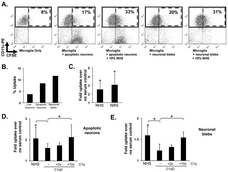
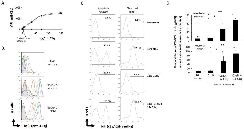
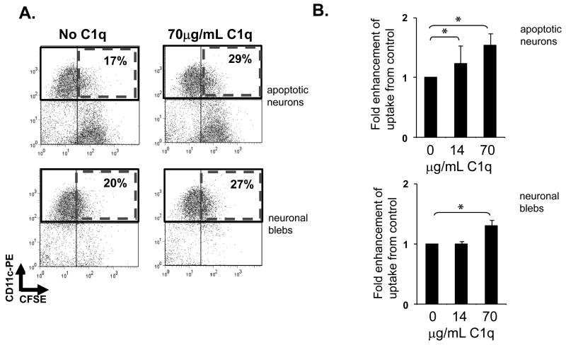
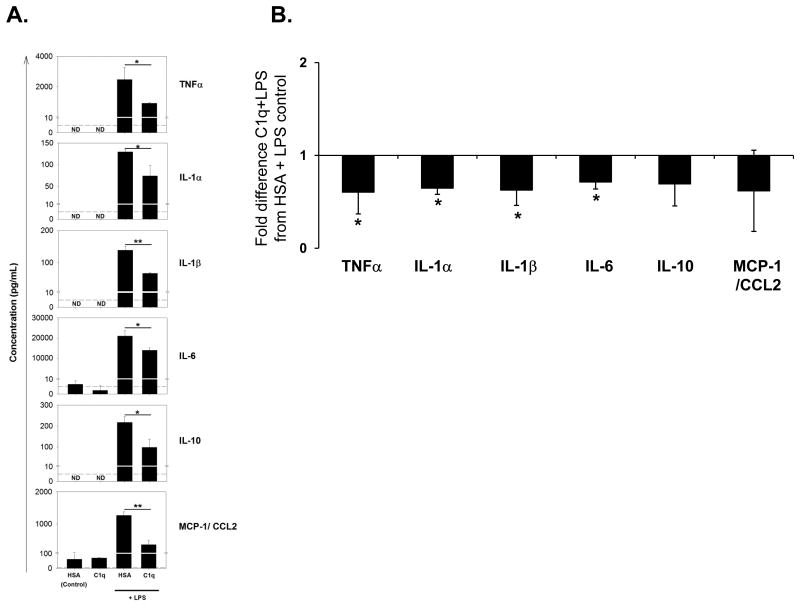
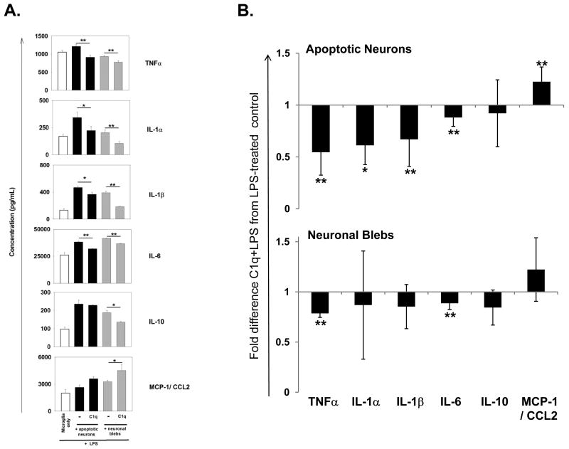
The expression of C1q, a recognition molecule of the complement system, is up-regulated following neuronal injury and is detected early in neurodegenerative disorders such as Alzheimer's disease. This multimeric protein triggers an enhancement of phagocytosis of suboptimally opsonized targets by microglia, the phagocytic cells of the CNS, similar to other phagocytes, enhances the uptake of apoptotic cells in peripheral phagocytes, and suppresses inflammatory cytokine production in human monocytes, macrophages and dendritic cells in the absence of activation of the entire complement cascade. The goal of this study was to determine if C1q could influence the inflammatory response to injury in the CNS, using primary rat microglia and neurons. The data show that microglia preferentially ingest apoptotic cells in comparison to live cells, like other professional phagocytes, that microglial ingestion of apoptotic neurons and neuronal blebs is enhanced by the presence of normal serum and that these enhanced levels of uptake are diminished in serum depleted of C1q. In addition, purified C1q bound to apoptotic neurons and neuronal blebs in a dose dependent manner, and alone triggered a significant enhancement of uptake by microglia. Microglia added to C1q coated wells or fed apoptotic neurons or neuronal blebs coated with C1q suppressed the lipopolysaccharide-induced production of proinflammatory cytokines interleukin (IL)-1alpha, IL-1beta, IL-6 and TNF-alpha, while the presence of C1q enhanced levels of the chemokine MCP-1/CCL2. The data are consistent with a protective role for C1q in the CNS during early stages of cell death by enhancing microglial clearance of apoptotic cells and suppressing proinflammatory cytokines.


Figures

Figure 1
Figure 1. Microglial uptake of apoptotic neurons and neuronal blebs
CFSE-labeled apoptotic neurons (A–D) and neuronal blebs (A,B,E) were added to rat microglia at a ratio of 3:1 (neurons) or 10:1 (neuronal blebs) without serum (B.) or with normal human serum (NHS), normal rat serum (NRS) (C), C1q-depleted human serum (C1qD) or C1qD in which C1q had been reconstituted to 2× or 10× serum concentration (D,E), as indicated, at a final volume of 10%. After incubation for 1 hour at 37°C, microglia were stained with anti CD11b and ingestion of CSFE targets was assessed by flow cytometry with representative dot blots from a single experiment shown in (A.). % uptake was assessed by calculation of the number of CD11b+CFSE+ cells as a % of total CD11b+ cells, with data from a single representative experiment in the absence of serum given in (B.). Average fold enhancement of uptake by sera are shown in (C. – E.) Data are expressed as the average fold enhancement of uptake over no serum control +/- SD. * p<0.05, ANOVA. n>5 (neurons), n=5 (blebs)
Figure 2
Figure 2. C1q and C3b binding to apoptotic neurons and neuronal blebs
Live neurons, apoptotic neurons and neuronal blebs were incubated with C1q or with normal human serum (NHS), C1q-depleted serum (C1qD) or C1qD in which C1q had been added to 2× or 10× serum concentration as described in Materials and Methods. A. C1q binding to live neurons (◆), apoptotic neurons (■) and neuronal blebs (□) incubated with amounts of human C1q from 7 to 300ug/mL, assessed by flow cytometry. MFI is calculated on the total population of cells B. Histogram depicting 0 (grey), 7 (red), 15 (orange), 75 (green), 150 (blue) or 300 (purple) μg/ml purified C1q binding to live neurons, apoptotic neurons or neuronal blebs assessed by flow cytometry. C. A typical histogram depicting the MFI of detection of anti- C3b/iC3b on apoptotic neurons (left) or neuronal blebs (right). Data in (A.), (B.) and (C.) are from a single experiment, representative of at least 3. D. Average C3b/iC3b binding (measured by MFI of antibody detection of C3d) expressed as % of the total average MFI of C3b/iC3b binding measured in samples incubated with 10% NHS. n=3-4 +/-SD. *p<0.05, **p<0.005, ANOVA.
Figure 3
Figure 3. Microglial uptake of apoptotic neurons and neuronal blebs
CFSE-labeled apoptotic neurons and neuronal blebs were added to rat microglia in the presence or absence of C1q as described in Materials and Methods. Uptake of CSFE-labeled targets by anti CD11b-labeled microglia was assessed by flow cytometry, with a single representative experiment depicted in (A) and expressed as average fold enhancement of uptake over control levels in the absence of C1q (B). +/- SD. * p<0.05 ANOVA, n=5 (apoptotic neurons), n=4 (neuronal blebs)
Figure 4
Figure 4. Modulation of LPS-induced proinflammatory cytokines released by rat microglia by C1q
Cytokine levels were measured by Luminex multiplex analysis of the supernatant of unstimulated microglia plated on LabTek chamber slides coated with 8 μg/mL C1q or HSA control, or identically treated cells stimulated with 150ng/mL LPS for 18 hours. (A.) Data shown are average cytokine levels from an individual experiment, measured in triplicate +/- SD. *p<0.05, **p<0.01, students t-test. Dashed line represents the lower limit threshold of detection. ND – not detected. (B.) Results are expressed as fold difference in expression in supernatants of microglia on C1q-coated slides and stimulated with LPS compared to control levels from cells incubated in wells coated with HSA and stimulated with LPS. Data are the average values of 3 separate experiments, each performed in triplicate +/- SD. * p<0.05, ANOVA.
Figure 5
Figure 5. C1q modulation of cytokines released from microglia during the uptake of apoptotic neurons and neuronal blebs
(A.) Cytokine levels were measured by Luminex multiplex analysis of the supernatant of microglia stimulated with 150 ng/ml LPS (white bar), or fed apoptotic neurons (black bars) or neuronal blebs (grey bars) in the absence or presence of 300 μg/mL C1q and stimulated with 150ng/mL LPS for 18 hours. Data are average cytokine levels from an individual experiment, measured in duplicate +/- SD. *p<0.05, **p<0.01, students t-test. (B.) Cytokine levels in the supernatant of LPS-stimulated microglia fed apoptotic neurons or neuronal blebs as measured in (A.), with results expressed as average fold difference in expression when C1q is present compared to control levels from identically treated microglia, in the absence of C1q, from n=5-7 individual experiments performed in duplicate with apoptotic neurons, or n=3-5 experiments with neuronal blebs) +/- SD. * p<0.05 **p<0.005, ANOVA.

References

    1. Bobak DA, Frank MM, Tenner AJ. C1q acts synergistically with phorbol dibutyrate to activate CR1-mediated phagocytosis by human mononuclear phagocytes. Eur J Immunol. 1988;18:2001–2007. - PubMed
    1. Botto M, Dell'agnola C, Bygrave AE, Thompson EM, Cook HT, Petry F, Loos M, Pandolfi PP, Walport MJ. Homozygous C1q deficiency causes glomerulonephritis associated with multiple apoptotic bodies. Nat Genet. 1998;19:56–59. - PubMed
    1. Botto M, Walport MJ. C1q, autoimmunity and apoptosis. Immunobiology. 2002;205:395–406. - PubMed
    1. Chen Y, Park YB, Patel E, Silverman GJ. IgM antibodies to apoptosis-associated determinants recruit C1q and enhance dendritic cell phagocytosis of apoptotic cells. J Immunol. 2009;182:6031–6043. - PMC - PubMed
    1. Chung EY, Liu J, Homma Y, Zhang Y, Brendolan A, Saggese M, Han J, Silverstein R, Selleri L, Ma X. Interleukin-10 Expression in Macrophages during Phagocytosis of Apoptotic Cells Is Mediated by Homeodomain Proteins Pbx1 and Prep-1. Immunity. 2007;27:952–964. - PMC - PubMed

Publication types

MeSH terms