. 2009;10(11):R129.
doi: 10.1186/gb-2009-10-11-r129. Epub 2009 Nov 17.

Differential binding and co-binding pattern of FOXA1 and FOXA3 and their relation to H3K4me3 in HepG2 cells revealed by ChIP-seq

Affiliations

Differential binding and co-binding pattern of FOXA1 and FOXA3 and their relation to H3K4me3 in HepG2 cells revealed by ChIP-seq

Mehdi Motallebipour et al. Genome Biol. 2009.

Abstract

Background: The forkhead box/winged helix family members FOXA1, FOXA2, and FOXA3 are of high importance in development and specification of the hepatic linage and the continued expression of liver-specific genes.

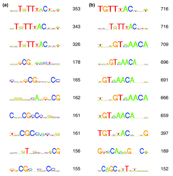
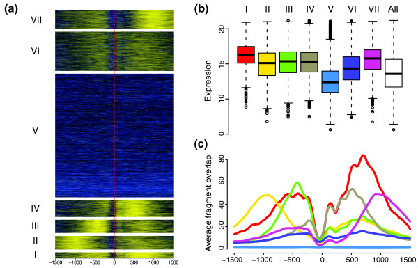
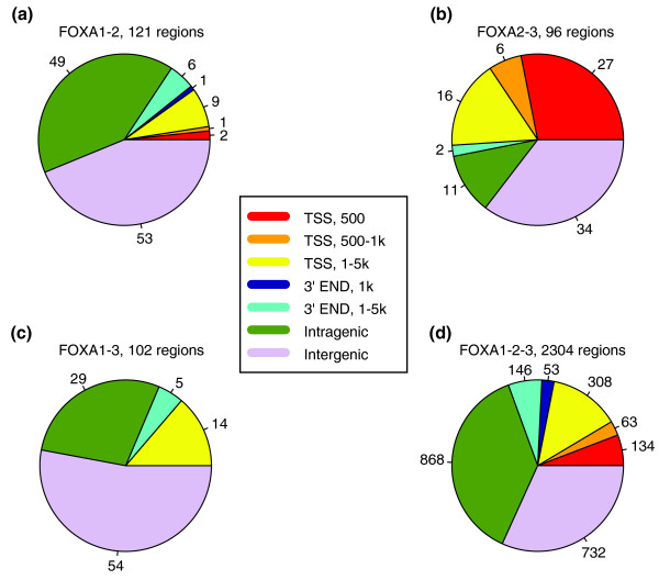
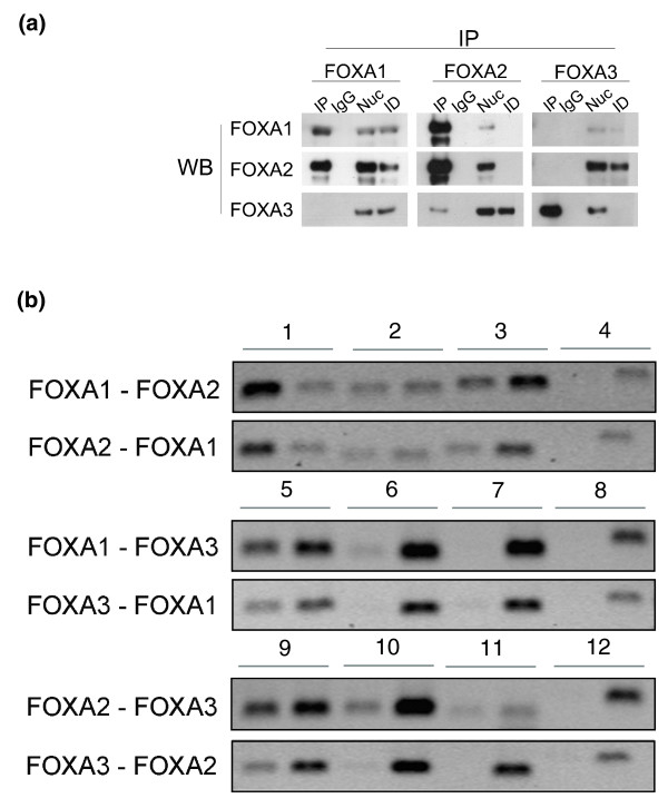
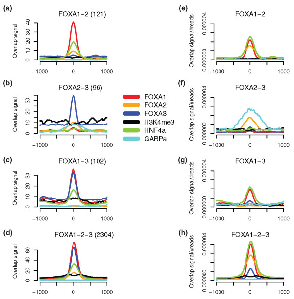
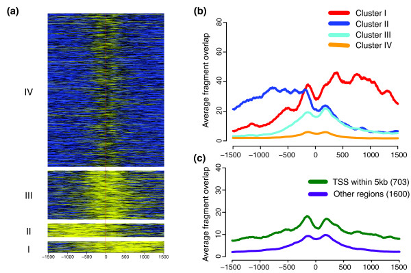
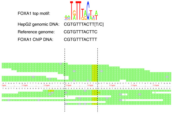
Results: Here, we present a genome-wide location analysis of FOXA1 and FOXA3 binding sites in HepG2 cells through chromatin immunoprecipitation with detection by sequencing (ChIP-seq) studies and compare these with our previous results on FOXA2. We found that these factors often bind close to each other in different combinations and consecutive immunoprecipitation of chromatin for one and then a second factor (ChIP-reChIP) shows that this occurs in the same cell and on the same DNA molecule, suggestive of molecular interactions. Using co-immunoprecipitation, we further show that FOXA2 interacts with both FOXA1 and FOXA3 in vivo, while FOXA1 and FOXA3 do not appear to interact. Additionally, we detected diverse patterns of trimethylation of lysine 4 on histone H3 (H3K4me3) at transcriptional start sites and directionality of this modification at FOXA binding sites. Using the sequence reads at polymorphic positions, we were able to predict allele specific binding for FOXA1, FOXA3, and H3K4me3. Finally, several SNPs associated with diseases and quantitative traits were located in the enriched regions.

Conclusions: We find that ChIP-seq can be used not only to create gene regulatory maps but also to predict molecular interactions and to inform on the mechanisms for common quantitative variation.


Figures

Figure 1
Figure 1
Results of de novo motif search. FOXA1 and FOXA3 data were analyzed using BCRANK as described in the Materials and methods. To the right of each motif is the assigned BCRANK score, which gives an indication of the quality of the motif. (a) Top ten predicted motifs for FOXA1. (b) Top ten predicted motifs for FOXA3.
Figure 2
Figure 2
H3K4me3 signals around the transcriptions start sites of 23,849 genes. (a) Enrichment of H3K4me3 in a window surrounding the TSSs. The genes were grouped into seven clusters (I to VII) by their H3K4me3 patterns as described in the Materials and methods section. The enrichment scale is from high (yellow) to low (blue), and the red vertical line represents the TSS position. Negative x-coordinates are upstream of the TSS and positive are downstream. (b) Box plots indicating the distributions of expression levels in the seven clusters. The white box represents the expression for all genes. (c) Average H3K4me3 signal footprints for the seven clusters. The colors are as in (b).
Figure 3
Figure 3
Genomic localization of common binding regions for FOXA1, FOXA2, and FOXA3. (a) FOXA1-2, (b) FOXA2-3, (c) FOXA1-3, and (d) FOXA1-2-3. Each region was mapped to all UCSC gene coordinates and sequentially matched to the categories 500 bp from TSS, 500 bp to 1 kb from TSS, 1 to 5 kb from TSS, 1 kb from 3'-end, 1 to 5 kb from 3'-end and intragenic. The intergenic group consists of those regions not matching any of the mentioned categories.
Figure 4
Figure 4
Co-immunoprecipitation and ChIP-reChIP of FOXAs reveals interaction and co-binding among FOXAs. (a) Immunoprecipitations were performed with indicated antibodies on nuclear extracts of HepG2 cells and the immunocomplexes were detected with FOXA1, FOXA2, and FOXA3 antibodies. IP, immunoprecipitation; IgG, the antibody was replaced by normal IgG; Nuc, total nuclear extract; ID, immunodepleted fraction obtained after IP. The blots are representative of two or three replicates. None of the proteins was overexpressed. (b) ChIP-reChIP of FOXA1, FOXA2, and FOXA3 tested by semiquantitative PCR. The order of antibodies used to immunoprecipitate the protein-DNA complex is indicated to the left. In each pair of bands, the left one is for the IP and the right for input. Pairs 1, 5, and 9: a primer amplifying a region with binding site for both proteins; pairs 2 and 6: regions with binding sites for FOXA1, but not FOXA2 or FOXA3, respectively; pairs 3 and 10: regions with binding sites for FOXA2, but not FOXA1 or FOXA3, respectively; pairs 7 and 11: regions with binding sites for FOXA3, but not FOXA1 or FOXA2, respectively; pairs 4, 8, and 12: a region with no FOXA binding. FOXA2-FOXA1, FOXA3-FOXA1, and FOXA3-FOXA2 were performed as independent experiments from the other three ChIP-reChIPs.
Figure 5
Figure 5
Enrichment signals in regions of pair-wise co-binding for FOXA1, FOXA2, and FOXA3. For each FOXA co-binding site, enrichment signals for FOXA1 (red), FOXA2 (orange), FOXA3 (blue), H3K4me3 (black), HNF4α (olive green), and GABP (turquoise) are plotted, centered on the putative FOXA binding site. (a-d) Graphs of the non-normalized data. (e-h) Graphs for each factor normalized to their number of aligned reads. Numbers in brackets for (a-d) are the number of sites with co-binding, as presented in Figure 3.
Figure 6
Figure 6
H3K4me3 signals around 2,303 FOXA1-2-3 regions. (a) Enrichment of H3K4me3 in a window surrounding the center of FOXA1-2-3 regions. The regions were grouped into four clusters (I to IV) by their H3K4me3 patterns. The enrichment scale is from high (yellow) to low (blue), and the red vertical line represents the FOXA1-2-3 centers. Negative x-coordinates are upstream of the centers and positive are downstream. (b) Average H3K4me3 signal footprints for the four clusters in (a). (c) Average H3K4me3 signal footprints for regions with a TSS within 5 kb independent of direction (green) and regions lacking a TSS within this distance (purple).
Figure 7
Figure 7
Preferential binding of FOXA1 at a heterozygous SNP. SNP rs7248104 is located in a FOXA1 binding sequence and FOXA1 is preferentially bound to one allele. At the top is the FOXA1 motif, predicted by the BCRANK method, followed by the sequence found in HepG2 with the alleles of the heterozygous SNP (T/C) in brackets. These are followed by the sequence in the reference genome and the sequence found in the FOXA1-reads. At the SNP position, the T-allele corresponds to the FOXA1 motif, which is found in all 15 FOXA1 reads, while the C-allele in the reference genome is not detected at all. The raw data for individual FOXA1 reads in the region are presented at the bottom, viewed in the SOLiD™ Alignment Browser tool. The positions marked in green correspond to bases (in the SOLiD™ two-base encoding) that align to the reference genome. In gray are the bases with a match error. The yellow bases correspond to positions with valid adjacent mismatches, indicating the locations of SNPs. The vertical hatched lines enclose the position of the motif.

References

    1. Kaestner KH, Knochel W, Martinez DE. Unified nomenclature for the winged helix/forkhead transcription factors. Genes Dev. 2000;14:142–146. - PubMed
    1. Friedman JR, Kaestner KH. The Foxa family of transcription factors in development and metabolism. Cell Mol Life Sci. 2006;63:2317–2328. doi: 10.1007/s00018-006-6095-6. - DOI - PMC - PubMed
    1. Carlsson P, Mahlapuu M. Forkhead transcription factors: key players in development and metabolism. Dev Biol. 2002;250:1–23. doi: 10.1006/dbio.2002.0780. - DOI - PubMed
    1. Lee CS, Friedman JR, Fulmer JT, Kaestner KH. The initiation of liver development is dependent on Foxa transcription factors. Nature. 2005;435:944–947. doi: 10.1038/nature03649. - DOI - PubMed
    1. Schrem H, Klempnauer J, Borlak J. Liver-enriched transcription factors in liver function and development. Part I: the hepatocyte nuclear factor network and liver-specific gene expression. Pharmacol Rev. 2002;54:129–158. doi: 10.1124/pr.54.1.129. - DOI - PubMed

Publication types

MeSH terms

LinkOut - more resources