Review
. 2009 Nov 17:7:97.
doi: 10.1186/1479-5876-7-97.

Inflammatory mechanisms in ischemic stroke: therapeutic approaches

Affiliations
Review

Inflammatory mechanisms in ischemic stroke: therapeutic approaches

Shaheen E Lakhan et al. J Transl Med. .

Abstract

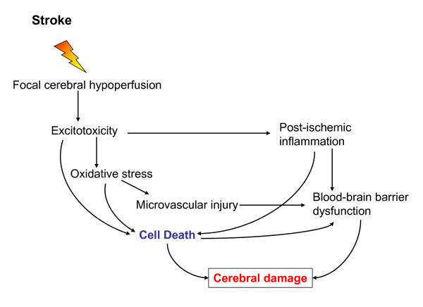
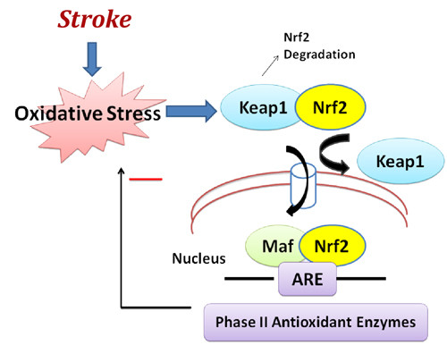
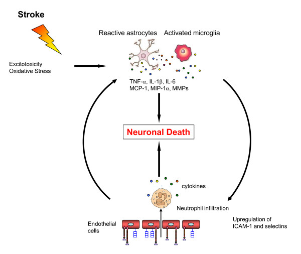
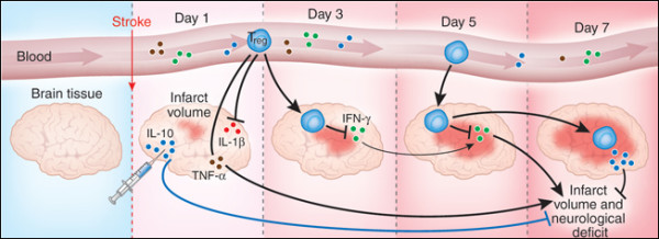
Acute ischemic stroke is the third leading cause of death in industrialized countries and the most frequent cause of permanent disability in adults worldwide. Despite advances in the understanding of the pathophysiology of cerebral ischemia, therapeutic options remain limited. Only recombinant tissue-plasminogen activator (rt-PA) for thrombolysis is currently approved for use in the treatment of this devastating disease. However, its use is limited by its short therapeutic window (three hours), complications derived essentially from the risk of hemorrhage, and the potential damage from reperfusion/ischemic injury. Two important pathophysiological mechanisms involved during ischemic stroke are oxidative stress and inflammation. Brain tissue is not well equipped with antioxidant defenses, so reactive oxygen species and other free radicals/oxidants, released by inflammatory cells, threaten tissue viability in the vicinity of the ischemic core. This review will discuss the molecular aspects of oxidative stress and inflammation in ischemic stroke and potential therapeutic strategies that target neuroinflammation and the innate immune system. Currently, little is known about endogenous counterregulatory immune mechanisms. However, recent studies showing that regulatory T cells are major cerebroprotective immunomodulators after stroke suggest that targeting the endogenous adaptive immune response may offer novel promising neuroprotectant therapies.


Figures

Figure 1
Figure 1
Ischemic cascade leading to cerebral damage. Ischemic stroke leads to hypoperfusion of a brain area that initiates a complex series of events. Excitotoxicity, oxidative stress, microvascular injury, blood-brain barrier dysfunction and postischemic inflammation lead ultimately to cell death of neurons, glia and endothelial cells. The degree and duration of ischemia determines the extent of cerebral damage.
Figure 2
Figure 2
Nuclear erythroid-related factor 2 (Nrf2) anti-oxidant signaling in acute ischemic stroke. Nrf2 is the principal transcription factor that regulates antioxidant response element (ARE)-mediated expression of phase II detoxifying antioxidant enzymes. Under normal conditions, Nrf2 is sequestered in the cytoplasm by an actin-binding (Kelch-like) protein (Keap1); on exposure of cells to oxidative stress, Nrf2 dissociates from Keap1, translocates into the nucleus, binds to ARE, and transactivates phase II detoxifying and antioxidant genes. Among the spectrum of antioxidant genes controlled by Nrf2 are catalase, superoxide dismutase (SOD), glutathione reductase, and glutathione peroxidase.
Figure 3
Figure 3
Postischemic inflammatory response. Excitotoxicity and oxidative stress caused by the initial ischemic event activate microglia and astrocytes which react by secreting cytokines, chemokines and matrix metalloproteases (MMP). These inflammatory mediators lead to an upregulation of cell adhesion molecules on endothelial cells, allowing blood-derived inflammatory cells, mainly neutrophils, to infiltrate the ischemic brain area. Neutrophils themselves also secrete cytokines which cause a further activation of glial cells. These processes all result in neuronal cell death and enhance the damage to the ischemic brain.
Figure 4
Figure 4
Regulatory T (Treg) cells protect the brain after stroke. Experiments by Liesz et al. [57] show that Treg cells prevent delayed lesion expansion in an IL-10-dependent manner in a mouse model of acute ischemic stroke. They also reduce the proinflammatory cytokine levels during the early postischemic inflammatory phase. Injection of IL-10 in the brain reduces infarct volume. Reprinted by permission from Macmillan Publishers Ltd: Nature Medicine 15, 138-139 Copyright 2009.

Similar articles

Cited by

References

    1. Lo EH, Dalkara T, Moskowitz MA. Mechanisms, challenges and opportunities in stroke. Nat Rev Neurosci. 2003;4:399–415. doi: 10.1038/nrn1106. - DOI - PubMed
    1. Donnan GA, Fisher M, Macleod M, Davis SM. Stroke. Lancet. 2008;371:1612–1623. doi: 10.1016/S0140-6736(08)60694-7. - DOI - PubMed
    1. American Heart Association. Heart Disease and Stroke Statistics - 2005 Update. Dallas, Texas: American Heart Association; 2005.
    1. Muir KW, Tyrrell P, Sattar N, Warburton E. Inflammation and ischaemic stroke. Curr Opin Neurol. 2007;20:334–342. doi: 10.1097/WCO.0b013e32813ba151. - DOI - PubMed
    1. Dirnagl U, Iadecola C, Moskowitz MA. Pathobiology of ischaemic stroke: an integrated view. Trends Neurosci. 1999;22:391–397. doi: 10.1016/S0166-2236(99)01401-0. - DOI - PubMed

MeSH terms