. 2010 Feb;113(2):358-66.
doi: 10.1093/toxsci/kfp281. Epub 2009 Nov 17.

Liver-specific ablation of integrin-linked kinase in mice results in enhanced and prolonged cell proliferation and hepatomegaly after phenobarbital administration

Affiliations

Liver-specific ablation of integrin-linked kinase in mice results in enhanced and prolonged cell proliferation and hepatomegaly after phenobarbital administration

Shashikiran Donthamsetty et al. Toxicol Sci. 2010 Feb.

Abstract

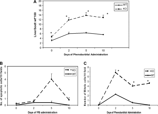
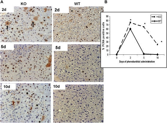
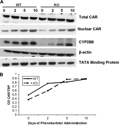
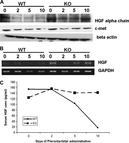
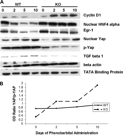
We have recently demonstrated that disruption of extracellular matrix (ECM)/integrin signaling via elimination of integrin-linked kinase (ILK) in hepatocytes interferes with signals leading to termination of liver regeneration. This study investigates the role of ILK in liver enlargement induced by phenobarbital (PB). Wild-type (WT) and ILK:liver-/- mice were given PB (0.1% in drinking water) for 10 days. Livers were harvested on 2, 5, and 10 days during PB administration. In the hepatocyte-specific ILK/liver-/- mice, the liver:body weight ratio was more than double as compared to 0 h at day 2 (2.5 times), while at days 5 and 10, it was enlarged three times. In the WT mice, the increase was as expected from previous literature (1.8 times) and seems to have leveled off after day 2. There were slightly increased proliferating cell nuclear antigen-positive cells in the ILK/liver-/- animals at day 2 as compared to WT after PB administration. In the WT animals, the proliferative response had come back to normal by days 5 and 10. Hepatocytes of the ILK/liver-/- mice continued to proliferate up until day 10. ILK/liver-/- mice also showed increased expression of key genes involved in hepatocyte proliferation at different time points during PB administration. In summary, ECM proteins communicate with the signaling machinery of dividing cells via ILK to regulate hepatocyte proliferation and termination of the proliferative response. Lack of ILK in the hepatocytes imparts prolonged proliferative response not only to stimuli related to liver regeneration but also to xenobiotic chemical mitogens, such as PB.


Figures

FIG. 1.
FIG. 1.
Quantitative assessment of hepatocyte proliferation, liver enlargement, and apoptosis. (A) Liver to body weight ratios of WT and ILK/liver−/− mice at days 0, 2 5, and 10 during PB administration. Each data point is the mean ± SE from more than three measurements per point. (B) Mitotic index as a marker of hepatocyte proliferation (C) apoptosis measured by terminal deoxynucleotidyl transferase–mediated deoxyuridine triphosphate nick-end labeling assay was estimated in WT and ILK KO mice at various days during PB. Mitotic and apoptotic cells were counted 10 different fields in four sections from four different knockout or control livers. While the apoptotic cells were counted at ×100 magnification, mitotic cells were counted at ×200 magnification. *Significantly different from the WT mice at that same time point (p ≤ 0.05).
FIG. 2.
FIG. 2.
(A) Representative photomicrographs of PCNA-stained liver section of ILK/liver−/− and WT mice at days 2, 5, and 10 during PB administration. Arrows show mitotic cells; magnification: ×200. (B) Hepatocyte proliferation was quantitated by counting the PCNA-positive cells. PCNA-positive cells were counted in low-power fields (×200) in four sections from four different knockout or control livers. Each data point is the mean ± SE from more than three measurements per point. *Significantly different from the WT mice at that same time point (p ≤ 0.05).
FIG. 3.
FIG. 3.
CAR activation in ILK/liver−/− and WT mice after PB administration. (A) Protein levels of CAR (nuclear and total cell extract) and CYP2B6 (total cell extract). Pooled liver samples from at least three mice were used for protein analysis. β-Actin- and TATA-binding protein were used as a loading control for total cell extract and nuclear extract, respectively. (B) Graphical representation of nuclear CAR expression following PB treatment. Expression levels were normalized to TATA-binding protein.
FIG. 4.
FIG. 4.
(A) HGF and its receptor cMet protein expression after PB administration in WT and ILK/liver−/− mice. Pooled liver samples from at least three mice were used for protein. β-Actin was used as the loading control. (B) Semiquantitative PCR for HGF in WT and ILK/liver−/− mice after PB administration. Pooled samples from at least three mice were for RNA isolation. (C) Serum HGF levels in WT and ILK/liver−/− mice before and after PB administration. Equal volumes of serum from three different animals were polled together for the analysis.
FIG. 5.
FIG. 5.
(A) Changes in cell cycle-related proteins in ILK/liver−/− and WT mice after PB administration. Pooled liver samples from at least three mice were used for protein. β-Actin and TATA-binding protein were used as loading controls for total cell extracts and nuclear extracts, respectively. (B) Graphical representation of YAP/p-YAP expression following PB treatment.

Similar articles

Cited by

References

    1. Apte U, Gkretsi V, Bowen WC, Mars WM, Luo JH, Donthamsetty S, Orr A, Monga SP, Wu C, Michalopoulos GK. Enhanced liver regeneration following changes induced by hepatocyte-specific genetic ablation of integrin-linked kinase. Hepatology. 2009;50:844–851. - PMC - PubMed
    1. Bell AW, Michalopoulos GK. Phenobarbital regulates nuclear expression of HNF-4alpha in mouse and rat hepatocytes independent of CAR and PXR. Hepatology. 2006;44:186–194. - PMC - PubMed
    1. Block GD, Locker J, Bowen WC, Petersen BE, Katyal S, Strom SC, Riley T, Howard TA, Michalopoulos GK. Population expansion, clonal growth, and specific differentiation patterns in primary cultures of hepatocytes induced by HGF/SF, EGF and TGF alpha in a chemically defined (HGM) medium. J. Cell Biol. 1996;132:1133–1149. - PMC - PubMed
    1. Burger PC, Herdson PB. Phenobarbital-induced fine structural changes in rat liver. Am. J. Pathol. 1966;48:793–809. - PMC - PubMed
    1. Carthew P, Edwards RE, Nolan BM. The quantitative distinction of hyperplasia from hypertrophy in hepatomegaly induced in the rat liver by phenobarbital. Toxicol. Sci. 1998;44:46–51. - PubMed

Publication types