. 2010 Jan;38(2):570-84.
doi: 10.1093/nar/gkp993. Epub 2009 Nov 17.

Yeast cohesin complex embraces 2 micron plasmid sisters in a tri-linked catenane complex

Affiliations

Yeast cohesin complex embraces 2 micron plasmid sisters in a tri-linked catenane complex

Santanu K Ghosh et al. Nucleic Acids Res. 2010 Jan.

Abstract

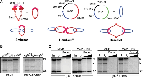
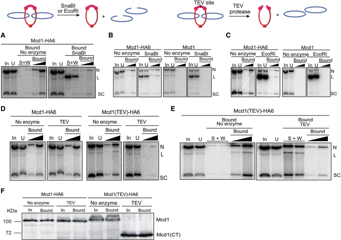
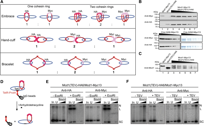
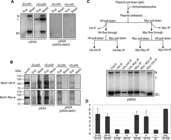
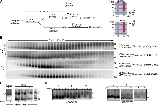
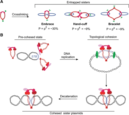
Sister chromatid cohesion, crucial for faithful segregation of replicated chromosomes in eukaryotes, is mediated by the multi-subunit protein complex cohesin. The Saccharomyces cerevisiae plasmid 2 micron circle mimics chromosomes in assembling cohesin at its partitioning locus. The plasmid is a multi-copy selfish DNA element that resides in the nucleus and propagates itself stably, presumably with assistance from cohesin. In metaphase cell lysates, or fractions enriched for their cohesed state by sedimentation, plasmid molecules are trapped topologically by the protein ring formed by cohesin. They can be released from cohesin's embrace either by linearizing the DNA or by cleaving a cohesin subunit. Assays using two distinctly tagged cohesin molecules argue against the hand-cuff (an associated pair of monomeric cohesin rings) or the bracelet (a dimeric cohesin ring) model as responsible for establishing plasmid cohesion. Our cumulative results most easily fit a model in which a single monomeric cohesin ring, rather than a series of such rings, conjoins a pair of sister plasmids. These features of plasmid cohesion account for its sister-to-sister mode of segregation by cohesin disassembly during anaphase. The mechanistic similarities of cohesion between mini-chromosome sisters and 2 micron plasmid sisters suggest a potential kinship between the plasmid partitioning locus and centromeres.


Figures

Figure 1.
Figure 1.
Topological models for cohesion; trapping an STB reporter plasmid in cohesin-associated form. (A) The subunits of the yeast cohesin complex, and the ring structure they are presumed to assemble, are schematically diagrammed. Shown next to it is a simplified representation of the cohesin ring used in figures to follow. The two STB reporter plasmids, pSG4 and pSG6, employed in these studies are symbolized by blue rings. One set of control assays made use of a derivative of pSG4 lacking the TetO21 sequence (pSG5). The three models for topological interaction between cohesin and sister plasmids tested in this study are shown. The hand-cuff is drawn to be consistent with the recent finding that dimerization of human cohesin is dependent on the Scc3 (SA1/SA2) subunit (25). (B) Following high-speed centrifugation of a cell lysate, DNA from the supernatant (Sup) and ‘chromatin’ pellet fractions, digested with EcoRI, was run in agarose gels and hybridized to a radiolabeled TRP1 probe. Results from a similar fractionation of a CEN4 minichromosome pTetO21CEN4 (20) are shown for comparison. Pl, plasmid; Ch, chromosome. (C) Cleared lysates (equivalent to ‘Sup’ in B) from metaphase [cir+] or [cir0] cells harboring pSG4 were immunoprecipitated with the HA-antibody and collected on Protein A beads. DNA extracted from the different fractions (In, input; U, unbound or flow-through) was probed by a radiolabeled fragment specific to pSG4. The amount of bound DNA loaded in the right lane was five times that in the left one. This ratio was kept constant in assays shown in subsequent figures as well. SC, supercoiled plasmid; L, linear plasmid; N, nicked plasmid.
Figure 2.
Figure 2.
Release of an STB reporter plasmid from cohesin’s grasp by linearizing DNA or cleaving Mcd1. The consequences of cutting DNA or protein on the topological association between plasmid and cohesin are schematically indicated. (A) Cohesin associated pSG4 was adsorbed on HA-antibody and immobilized on Protein A beads as described under Figure 1. After digestion with SnaBI, DNA released into the supernatant (S) and wash fractions (W) and that retained on the beads (Bound) were analyzed. (B and C) SnaBI or EcoRI digestion was performed in the cleared lysates prior to immunoprecipitation by HA-antibody. (D) Cleared lysates were treated with TEV protease, and then subjected to pull-down by HA-antibody and Protein A beads. (E) Cohesin bound plasmid from cleared lysates was treated with HA-antibody, trapped on Protein A beads, and subjected to TEV protease treatment. (F) Cleavage of Mcd1-HA6 by TEV protease in the cleared lysates (corresponding to the DNA analysis shown in D) was monitored by western blotting using HA-antibody. The amount of protein analyzed from the bead-bound fraction was four equivalents of that from the input. The identity of the weak band above Mcd1 seen in some of the lanes is not known. Its mobility would be consistent with phosphorylation of Mcd1, which occurs during the establishment of cohesion in response to DNA damage (27).
Figure 3.
Figure 3.
Plasmid–cohesin association in metaphase cells expressing two differentially tagged cohesin moieties. (A) The types of sister plasmid cohesion expected from the embrace, hand-cuff and bracelet models in presence of cohesin(Mcd1-HA6) and cohesin(Mcd1-Myc13) in equivalent amounts are indicated (see also Table 1). Whereas Mcd1-HA6 in cohesin could be cleaved by TEV protease, Mcd1-Myc13 could not. (B) Aliquots of cell lysates were probed by western analysis using HA- or Myc-antibody to reveal relative levels of cohesin(Mcd1-HA6) or cohesin(Mcd1-Myc13). Data are shown for the haploid strains expressing either cohesin(Mcd1-HA6) (lane 1) or cohesin(Mcd1-Myc13) (lane 2) and the diploid generated from them expressing both Mcd1 variants (lanes 3–7). Signals from the two antibodies were normalized using aliquots of ∼75% pure Cre recombinase tagged at its carboxyl-terminus with HA6 as well as Myc13. The mean Myc13 to HA6 signal intensity was 1.83 ± 0.18. The dilution factor between Cre samples stained by Coomassie blue (right) and the corresponding ones analyzed by western blotting (left) was 500 to 1. (C) Aliquots of cell lysates from the haploid and diploid strains, run as in B, were probed using an antibody to native Mcd1. The mean ratio of Mcd1-Myc13 to Mcd1-HA6 was 0.96 ± 0.11. (D) pSG4 molecules from the cleared lysate were first immobilized on IgG beads, and then released from them by disrupting TetO–TetR interaction using anhydrotetracycline. (E and F) Following treatment with EcoRI or TEV protease, plasmid pull-down was attempted using HA- or Myc-antibody.
Figure 4.
Figure 4.
Distinction between embrace and bracelet models for plasmid cohesion: cohesin stoichiometry tested by sequential immunoprecipitation. (A and B) The expected outcomes for plasmid immobilization via TetO affinity interaction followed by DNA linearization were experimentally verified. Plasmid molecules associated with Protein A-TetR bound to TetO were pulled down by IgG beads. DNA and protein remaining associated with the beads or released into the supernatant in the absence of EcoRI treatment or following EcoRI digestion were followed by Southern and western analyses, respectively. (C) The flow-chart for the two-step immunoprecipitation assays is diagrammed at the top. Plasmids were first trapped on IgG beads as in A, and then released by treatment with anhydrotetracycline. Equal amounts of the supernatant containing the freed plasmid were immunoprecipitated with the HA- or Myc-antibody. The leftover plasmid molecules in the supernatant were subjected to a second round of immunoprecipitations. (D) The histograms denote the mean ratios of Southern blot signals for immunoprecipitated DNA from three independent experiments, with the error bars showing standard deviations. Immunoprecipitations with HA- and Myc-antibodies are represented by ‘H’ and ‘M’, respectively. Sequential immunoprecipitations by these antibodies are indicated by the two letters separated by a dash. The ratio of the input (IN) plasmid DNA to that immunoprecipitated by the HA- and Myc-antibodies combined is given as IN/[H + M]. The immunoprecipitable plasmid fractions were 17.33%, 16.50%, 16.46% in individual assays.
Figure 5.
Figure 5.
Enrichment of plasmids in their cohesed form from metaphase cells by velocity sedimentation: test of the topological model for cohesion. (A) The experimental regimen for enriching metaphase cells from the appropriate [cir0] host strain harboring pSG6 and going through the cell cycle in glucose or galactose is schematically indicated. At 45 min for the cell cycle in glucose and at 75 min for that in galactose, the predominant population consisted of large budded cells with a single DAPI staining mass at the bud neck. (B) The sedimentation patterns of pSG6 in 12.5–37.5% sucrose gradient were followed under conditions where the plasmid-borne CEN alone or STB alone or neither of the two was active. Samples were run in agarose gels in the cold (4°C) and probed by pSG6-specific radio-labeled DNA. C, cohesed plasmids; NC, non-cohesed plasmids. (C) Representative fast (F), intermediate (I) and slow (S) sedimenting fractions from the gradient were reanalyzed by agarose gel electrophoresis with or without EcoRI digestion, followed by SDS treatment. For reference, S fractions treated or untreated with EcoRI but without subsequent SDS addition (left panel) and DNA prepared form the lysate by phenol extraction and ethanol precipitation (rightmost lane) were included in the run. (D and E) Representative fractions from the sucrose gradient (fast, slow and intermediate) were treated with EcoRI (D) or with TEV protease (E), and subjected to agarose gel electrophoresis under native conditions.
Figure 6.
Figure 6.
A single ring formed by a monomeric cohesin complex as the unit of cohesion at STB. (A) The results from Haering et al. (18) for CEN cohesion ruling out ‘double’ rings of cohesin are schematically represented. In their experimental design, covalent protein ring closure required crosslinking two neighboring pairs of cysteines at two locations (green circles) to form a pair of chemical bridges (green triangles). Cross-linking efficiency (or probability ‘p’ of circle to triangle conversion) was ∼55% in their assays. Experiments agreed with entrapment probability of DNA sisters ‘P’ equal to p2 (30%) and not p4 (9%). Note that, upon SDS treatment during the assay, a physical hand-cuff (but not a topological one) would fall apart to yield monomeric cohesin rings, each with a single trapped plasmid molecule. (B) In the pre-cohesed state of the 2 micron circle, multiple cohesin molecules may interact physically and dynamically at or near STB. Such interactions could be promoted by the cohesin loading factors Scc2 and Scc4, which are required for cohesin assembly on the plasmid (5). Transition to the stable topological association may be mediated by passage of the replisome and closure of a single cohesin ring around a pair of STB sisters.

Similar articles

Cited by

References

    1. Nasmyth K, Schleiffer A. From a single double helix to paired double helices and back. Phil. Trans. R Soc. Lond. B Biol. Sci. 2004;359:99–108. - PMC - PubMed
    1. Onn I, Heidinger-Pauli JM, Guacci V, Unal E, Koshland DE. Sister chromatid cohesion: a simple concept with a complex reality. Annu. Rev. Cell Dev. Biol. 2008;24:105–129. - PubMed
    1. Uhlmann F. The mechanism of sister chromatid cohesion. Exp. Cell Res. 2004;296:80–85. - PubMed
    1. Watanabe Y. Sister chromatid cohesion along arms and at centromeres. Trends Genet. 2005;21:405–412. - PubMed
    1. Mehta S, Yang XM, Chan CS, Dobson MJ, Jayaram M, Velmurugan S. The 2 micron plasmid purloins the yeast cohesin complex: a mechanism for coupling plasmid partitioning and chromosome segregation? J. Cell Biol. 2002;158:625–637. - PMC - PubMed

Publication types

MeSH terms