. 2010 Jan 22;285(4):2676-85.
doi: 10.1074/jbc.M109.043471. Epub 2009 Nov 17.

Estrogen-mediated regulation of Igf1 transcription and uterine growth involves direct binding of estrogen receptor alpha to estrogen-responsive elements

Affiliations

Estrogen-mediated regulation of Igf1 transcription and uterine growth involves direct binding of estrogen receptor alpha to estrogen-responsive elements

Sylvia C Hewitt et al. J Biol Chem. .

Abstract

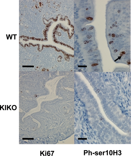
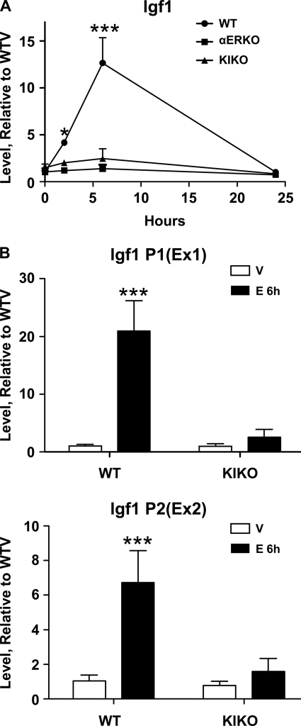
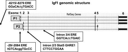
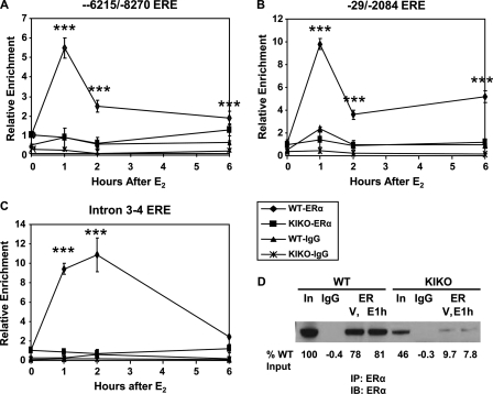
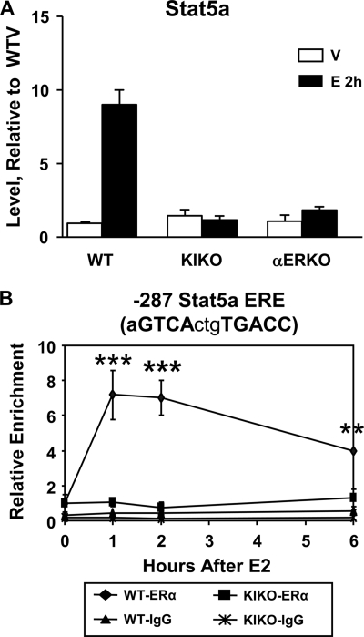
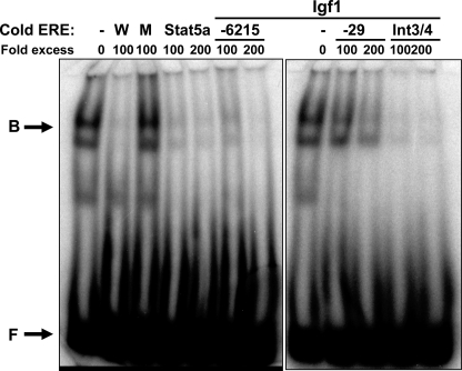
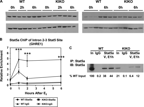
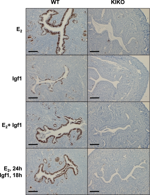
Estrogen enables uterine proliferation, which depends on synthesis of the IGF1 growth factor. This proliferation and IGF1 synthesis requires the estrogen receptor (ER), which binds directly to target DNA sequences (estrogen-responsive elements or EREs), or interacts with other transcription factors, such as AP1, to impact transcription. We observe neither uterine growth nor an increase in Igf1 transcript in a mouse with a DNA-binding mutated ER alpha (KIKO), indicating that both Igf1 regulation and uterine proliferation require the DNA binding function of the ER. We identified several potential EREs in the Igf1 gene, and chromatin immunoprecipitation analysis revealed ER alpha binding to these EREs in wild type but not KIKO chromatin. STAT5 is also reported to regulate Igf1; uterine Stat5a transcript is increased by estradiol (E(2)), but not in KIKO or alpha ERKO uteri, indicating ER alpha- and ERE-dependent regulation. ER alpha binds to a potential Stat5a ERE. We hypothesize that E(2) increases Stat5a transcript through ERE binding; that ER alpha, either alone or together with STAT5, then acts to increase Igf1 transcription; and that the resulting lack of IGF1 impairs KIKO uterine growth. Treatment with exogenous IGF1, alone or in combination with E(2), induces proliferation in wild type but not KIKO uteri, indicating that IGF1 replacement does not rescue the KIKO proliferative response. Together, these observations suggest in contrast to previous in vitro studies of IGF-1 regulation involving AP1 motifs that direct ER alpha-DNA interaction is required to increase Igf1 transcription. Additionally, full ER alpha function is needed to mediate other cellular signals of the growth factor for uterine growth.


Figures

FIGURE 1.
FIGURE 1.
E2 does not increase uterine proliferation markers Ki67 or phosphoserine 10 histone H3 in the KIKO. Uterine cross-sections from ovariectomized mice that were treated for 24 h with E2, representative of three to five uteri sampled were incubated with anti-Ki67 or anti-Ser(P)10 histone H3 (Ph-ser10H3) as markers of cell cycle activity. Only the WT epithelia had easily detectable amounts of either marker. The bar in the Ki67 section is 0.1 μm, and the bar in the anti-Ser(P)10 histone H3 section is 0.2 μm. The arrow shows a perimitotic epithelial cell.
FIGURE 2.
FIGURE 2.
A, Igf1 transcript is not induced by E2 in the KIKO or αERKO uterus. Total uterine RNA was isolated from uteri from ovariectomized WT, KIKO, or αERKO mice that had been treated with vehicle (0 h) or with E2 for 2, 6, or 24 h. Transcripts were quantified by real time PCR as described under “Materials and Methods” and calculated relative to WT vehicle-treated (WTV). WT, KIKO, and αERKO E2 treated were tested versus their vehicle control by two-way ANOVA with Bonferroni correction (*, p < 0.05; ***, p < 0.001) in GraphPad Prism software (GraphPad Software Inc, La Jolla, CA). B, Igf1 transcripts from both promoter 1 and 2 are increased by E2. Total RNA from uteri treated with vehicle or with E2 for 6 h was quantified by real time RT-PCR using primers that are specific for exons 1 or 2, reflecting use of promoter 1 or 2 (P1 and P2), respectively. Both transcripts are increased by E2 and are not induced in the KIKO. The data were tested by two-way ANOVA t test with a Bonferroni correction (***, p < 0.001) compared with vehicle control.
FIGURE 3.
FIGURE 3.
Igf1 gene structure. The mouse Igf1 gene consists of six exons. According to RefSeq, five different splice variants have been confirmed. Two different promoters (P1 and P2) result in selective use of exon 1 or 2 as a first exon. Sequence analysis has identified a potential ERE site −8270 or −6215 base pairs upstream of promoters 2 and 1, respectively. Additional potential EREs are located adjacent to exon 1 (−29 bp/−2084 bp) and in the large 48,000 bp intron between exons 3 and 4. Stat5-binding sites (GHRE1) have been described in the intron between exons 2 and 3.
FIGURE 4.
FIGURE 4.
ERα is recruited to ERE sequences after E2 treatment. ChIP was carried out on chromatin isolated from WT or KIKO uteri after ovariectomy and injection with vehicle (0 h) or E2 for 1, 2, or 6 h. A–C, chromatin immunoprecipitated with anti-ERα antibody or normal IgG was analyzed by real time PCR using primers that flanked the ERE sequence identified: upstream of both promoters 1 and 2 (−6215 and 8270 bp, respectively) (A); adjacent to exon 1 (−29/−208 4bp) (B); or the ERE sequence in intron 3–4 (C). The levels are expressed relative to the values for WT vehicle anti-ERα samples, which are plotted as time 0. The time 0 ERα value was compared with each time point ERα value by two-way ANOVA t test with Bonferroni correction. For each time point, p < 0.001 (***). D, ERα Western blot of input (In) and chromatin-associated proteins in ChIP sample aliquots after IP with normal IgG control (IgG) or anti-ERα. Samples were from WT or KIKO treated with saline vehicle (V) or treated for 1 h with E2 (E1h). ERα antibody: sc542, Santa Cruz diluted 1:1000 in TBST with 5% milk. Gel images were quantified using ImageQuant software (GE Healthcare), and the values were calculated as percentages of WT input, shown below the gel. IP, immunoprecipitation; IB, immunoblot.
FIGURE 5.
FIGURE 5.
Estrogen regulation of Stat5a. A, RT-PCR analysis for Stat5a transcript in WT or KIKO and αERKO uterine samples treated for 2 h with vehicle or E2. The data were tested by two-way ANOVA t tests with a Bonferroni correction. ***, p < 0.001 compared with vehicle control. B, ChIP analysis of an ERE sequence in the Stat5a promoter. ChIP was carried out on uterine chromatin isolated after injection with vehicle or E2 for 1, 2, or 6 h. Immunoprecipitation was done with anti-ERα antibody or normal IgG. DNA was analyzed by real time PCR using primers that flanked the ERE sequence 287 bp upstream of the Stat5a transcript. The levels are expressed relative to the values for WT vehicle anti-ERα samples, which are plotted as time 0. The time 0 ERα value was compared with each time point ERα value by two-way ANOVA t tests with Bonferroni correction. For each time point p < 0.001 (***) or p < 0.01 (**).
FIGURE 6.
FIGURE 6.
Gel shift demonstrates Stat5a and Igf1 ERE sequences bind to ERα. MCF-7 nuclear proteins were bound with 32P-labeled WT consensus ERE sequence and competed with 100- or 200-fold excess of unlabeled WT ERE (W) ERE mutated so it does not bind ERα (M) or the ERE sequences from Stat5a, or from Igf1 −6215. −29, or intron 3–4. The doublet of ERα-bound probe (B) or free probe (F) are indicated by arrows.
FIGURE 7.
FIGURE 7.
E2 does not increase STAT5a protein but causes accumulation in the nuclear fraction. A, Western blots from total uterine proteins (left panels) or nuclear extracts (right panels) from WT or KIKO mice treated with vehicle (0 h) or with E2 for 2 or 6 h detected with anti-STAT5a (top panels, both left and right; Santa Cruz, sc1081 diluted 1:1000 in TBST with 5% Milk). β-Tubulin (bottom left panel; Santa Cruz sc-9104 diluted 1:2000 in TBTS and milk) or β-actin (bottom right panel; Santa Cruz sc1616 diluted 1:5000 in TBST and milk) is the loading control. B, ChIP analysis of STAT5-binding site in intron 2–3 of the Igf1 gene. Chromatin isolated from mice treated with vehicle or E2 for 1, 2, or 6 h was immunoprecipitated with anti-Stat5a or normal IgG. Precipitated DNA was quantified by real time PCR using primers that flanked the GHRE-1 site. The levels are expressed relative to the values for WT vehicle anti-STAT5a or sample, which are plotted as time 0. The time 0 Stat5a value was compared with each time point Stat5a value by two-way ANOVA t tests with Bonferroni correction. For each time point p < 0.001 (***). C, Western blot of input (In) and chromatin associated proteins in ChIP sample aliquots after IP with normal IgG control (IgG) or anti-STAT5a and blotted as in A. The samples were from WT or KIKO treated with saline vehicle (V) or for 1 h with E2 (E1h). The gel images were quantified using ImageQuant software (GE Healthcare), and the values were calculated as percentages of WT input, shown below the gel. IP, immunoprecipitation; IB, immunoblot.
FIGURE 8.
FIGURE 8.
Exogenous Igf1 does not rescue KIKO epithelial proliferation. Ovariectomized WT or KIKO mice were treated with E2, longR3 Igf1, or both. Either they were treated simultaneously, or Igf1 was administered 6 h after the E2 injection to mimic the peak of Igf1 induction that follows E2 stimulation. Uterine tissue was collected after 24 h, and cross-sections were evaluated for the cell proliferation marker Ki67. The bar represents 0.1 μm; the sections are representative of the treatment group, which included three mice each.

Similar articles

Cited by

References

    1. Shao W., Brown M. (2004) Breast Cancer Res. 6, 39–52 - PMC - PubMed
    1. Jordan V. C. (2007) Nat Rev Cancer 7, 46–53 - PubMed
    1. Safe S., Kim K., Kim K. (2008) J. Mol. Endocrinol. 41, 263–275 - PMC - PubMed
    1. Lahooti H., White R., Danielian P. S., Parker M. G. (1994) Mol. Endocrinol. 8, 182–188 - PubMed
    1. Jakacka M., Ito M., Weiss J., Chien P. Y., Gehm B. D., Jameson J. L. (2001) J. Biol. Chem. 276, 13615–13621 - PubMed

Publication types

MeSH terms

LinkOut - more resources