. 2010 Jan 15;285(3):2193-202.
doi: 10.1074/jbc.M109.058248. Epub 2009 Nov 17.

The FGFRL1 receptor is shed from cell membranes, binds fibroblast growth factors (FGFs), and antagonizes FGF signaling in Xenopus embryos

Affiliations

The FGFRL1 receptor is shed from cell membranes, binds fibroblast growth factors (FGFs), and antagonizes FGF signaling in Xenopus embryos

Florian Steinberg et al. J Biol Chem. .

Abstract

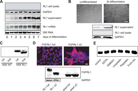
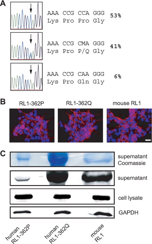
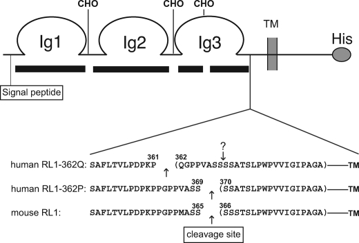
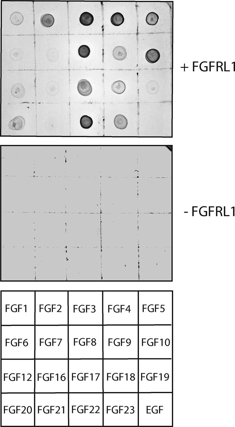
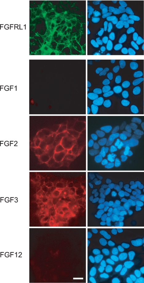
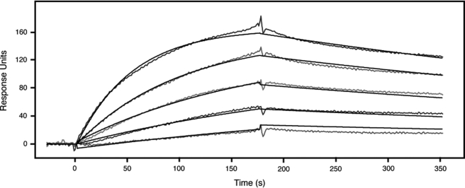
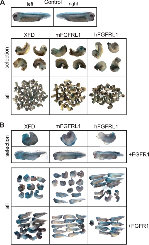
FGFRL1 (fibroblast growth factor receptor like 1) is the fifth and most recently discovered member of the fibroblast growth factor receptor (FGFR) family. With up to 50% amino acid similarity, its extracellular domain closely resembles that of the four conventional FGFRs. Its intracellular domain, however, lacks the split tyrosine kinase domain needed for FGF-mediated signal transduction. During embryogenesis of the mouse, FGFRL1 is essential for the development of parts of the skeleton, the diaphragm muscle, the heart, and the metanephric kidney. Since its discovery, it has been hypothesized that FGFRL1 might act as a decoy receptor for FGF ligands. Here we present several lines of evidence that support this notion. We demonstrate that the FGFRL1 ectodomain is shed from the cell membrane of differentiating C2C12 myoblasts and from HEK293 cells by an as yet unidentified protease, which cuts the receptor in the membrane-proximal region. As determined by ligand dot blot analysis, cell-based binding assays, and surface plasmon resonance analysis, the soluble FGFRL1 ectodomain as well as the membrane-bound receptor are capable of binding to some FGF ligands with high affinity, including FGF2, FGF3, FGF4, FGF8, FGF10, and FGF22. We furthermore show that ectopic expression of FGFRL1 in Xenopus embryos antagonizes FGFR signaling during early development. Taken together, our data provide strong evidence that FGFRL1 is indeed a decoy receptor for FGFs.


Figures

FIGURE 1.
FIGURE 1.
FGFRL1 is shed from the surface of C2C12 and HEK293 cells. A, C2C12 myogenic stem cells were induced to differentiate into myotubes by serum withdrawal over 7 days of culture. Northern blot analysis of FGFRL1 mRNA (bottom panel) and Western blot detection of FGFRL1 protein in lysates of differentiating C2C12 myoblasts (top panel) and in the corresponding supernatants (middle panel). B, primary myoblasts obtained from tongue muscle are shown before and after 3 days of in vitro differentiation (top panels; bar, 100 μm). The Western blots (bottom panel) show that undifferentiated, proliferating myoblasts (left top panel) secrete very little FGFRL1, whereas differentiating myoblasts (right top panel) released a larger amount of FGFRL1 into the cell culture medium over 3 days of culture. No FGFRL1 could be detected in the cell lysates of myoblasts and myotubes. C, Western blot analysis of FGFRL1 in the cell culture medium (sup) and in the cell lysate (lys) of HEK293 wild type (WT) cells and HEK293 cells overexpressing FGFRL1 (HEK RL1). Half of purified supernatant and 120 of the lysed cell layers was loaded onto the gel. D, stable overexpression of full-length (RL1 full) and C-terminally truncated FGFRL1 (RL1 ΔC) in HEK293 cells. The immunofluorescence analysis (upper panels) shows that the full-length receptor resides mostly in intracellular vesicles, whereas RL1ΔC accumulates at the cell membrane. Bar, 20 μm. Western blot (bottom panel) detection of FGFRL1 in cell lysates (left top panel) and in the supernatants (right top panel). Note that both proteins were shed in equal amounts. E, protease inhibitor treatment of stably transfected HEK293 cells shedding FGFRL1. The cells were incubated in serum-free medium containing the indicated inhibitors for 24 h. The supernatants were then heparin-purified, and the eluates were subjected to Western blot detection of FGFRL1. None of the inhibitors had any effect on the shedding of FGFRL1.
FIGURE 2.
FIGURE 2.
A polymorphism in the membrane-proximal region affects the shedding of FGFRL1 from HEK293 cells. A, genomic sequencing of exon 6 of the FGFRL1 gene from 96 healthy British donors. A single nucleotide polymorphism results in the exchange of proline 362 in the membrane-proximal region to glutamine. The percentages of individuals carrying the respective alleles are shown. B, stable, cytomegalovirus promoter-driven overexpression of C-terminally deleted FGFRL1 constructs corresponding to the RL1–362P (left panel) and the RL1–362Q (middle panel) allele as well as the murine protein (right panel). No obvious differences in expression levels or subcellular distribution were detected. Bar, 20 μm. C, Western blot analysis of the three proteins in cell layers and in cell culture supernatants. No difference in overall expression in the cell layer was detected (bottom panel). Significantly more FGFRL1 was purified from supernatants of the RL1–362Q expressing cells, indicating that the shedding of this FGFRL1 variant is enhanced (middle panels).
FIGURE 3.
FIGURE 3.
Mass spectrometric analysis of the cleavage sites. A schematic representation of the FGFRL1 receptor is shown. The black bars beneath the scheme indicate the sequence coverage of the FGFRL1 ectodomain, which was 94% of the nonglycosylated peptides. The gaps in coverage most likely represent sites of N-glycosylation, indicated by CHO. Shown below the receptor scheme are the most C-terminal peptides that were identified for the human RL1–362Q and RL1–362P receptors and the mouse FGFRL1. Arrows indicate the proteolytic cleavage sites. Note that the exchange of proline 362 by glutamine results in a shift of the cleavage site to the polymorphic glutamine residue. The original cleavage site is still present in the RL1–362Q protein and is indicated by an arrow with a question mark.
FIGURE 4.
FIGURE 4.
Ligand dot blot analysis reveals differential FGF binding preferences of human FGFRL1. Soluble, Myc-tagged FGFRL1 was used to probe a blot of spotted recombinant FGFs (200 ng each). In the control blot, Myc-tagged FGFRL1 was omitted. The bottom panel shows the positions of the spotted FGFs and the control epidermal growth factor on the blot.
FIGURE 5.
FIGURE 5.
A fluorescent binding analysis on living cells reveals differential FGF binding preferences of human FGFRL1. HEK293 cells expressing FGFRL1 were incubated with selected fluorescently labeled FGFs (DyLight 547, red channel). The subcellular localization of FGFRL1 (Cy-2, green channel) is shown in the top panel. The right panels show the cell nuclei after staining with 4′,6′-diamino-2-phenylindole. Note that FGF2 and FGF3 bind to the surface of the FGFRL1 expressing cells, whereas FGF1 and FGF12 do not bind. Bar, 20 μm.
FIGURE 6.
FIGURE 6.
Surface plasmon resonance analysis of the binding of FGFRL1 to FGF3. FGF3 was coupled to a Biacore sensor chip and increasing concentrations (3.13, 6.25, 12.5, 25, and 50 nm) of FGFRL1 ectodomain were injected over the sensor chip. The binding and dissociation kinetics shown in the diagram were integrated by the Biacore analysis software and resulted in a dissociation constant (Kd) of 4.16 × 10⁻⁹ m.
FIGURE 7.
FIGURE 7.
FGFRL1 antagonizes FGF signaling in Xenopus development. Both blastomeres of two-cell stage embryos were coinjected with XFD, mouse FGFRL1 (mFGRL1), or human FGFRL1 (hFGFRL1) mRNA (300 pg/blastomere) and 250 pg of mRNA for the lineage tracer nuclear β-galactosidase. Control embryos injected with lineage tracer only were used to determine stage 35/36 as the end point of development, where all embryos were fixed and processed for β-galactosidase activity. All of the injected embryos are shown beside selected examples presented in close-up views. A, left and right sides of injected control embryos develop normally (top panel). Embryos injected with XFD, mouse FGFRL1, or human FGFRL1 display similar phenotypes (bottom panel). B, coinjection of FGFR1 mRNA together with XFD, mFGFRL1, or hFGFRL1 mRNA rescues the XFD phenotype.

Similar articles

Cited by

References

    1. Beenken A., Mohammadi M. (2009) Nat. Rev. Drug Discov. 8, 235–253 - PMC - PubMed
    1. Itoh N., Ornitz D. M. (2004) Trends Genet. 20, 563–569 - PubMed
    1. Itoh N., Ornitz D. M. (2008) Dev. Dyn 237, 18–27 - PubMed
    1. Wiedemann M., Trueb B. (2000) Genomics 69, 275–279 - PubMed
    1. Sleeman M., Fraser J., McDonald M., Yuan S., White D., Grandison P., Kumble K., Watson J. D., Murison J. G. (2001) Gene 271, 171–182 - PubMed

Publication types

MeSH terms