. 2010 Jan 15;285(3):1754-64.
doi: 10.1074/jbc.M109.063826. Epub 2009 Nov 17.

Pin1 promotes transforming growth factor-beta-induced migration and invasion

Affiliations

Pin1 promotes transforming growth factor-beta-induced migration and invasion

Isao Matsuura et al. J Biol Chem. .

Abstract

Transforming growth factor-beta (TGF-beta) regulates a wide variety of biological activities. It induces potent growth-inhibitory responses in normal cells but promotes migration and invasion of cancer cells. Smads mediate the TGF-beta responses. TGF-beta binding to the cell surface receptors leads to the phosphorylation of Smad2/3 in their C terminus as well as in the proline-rich linker region. The serine/threonine phosphorylation sites in the linker region are followed by the proline residue. Pin1, a peptidyl-prolyl cis/trans isomerase, recognizes phosphorylated serine/threonine-proline motifs. Here we show that Smad2/3 interacts with Pin1 in a TGF-beta-dependent manner. We further show that the phosphorylated threonine 179-proline motif in the Smad3 linker region is the major binding site for Pin1. Although epidermal growth factor also induces phosphorylation of threonine 179 and other residues in the Smad3 linker region the same as TGF-beta, Pin1 is unable to bind to the epidermal growth factor-stimulated Smad3. Further analysis suggests that phosphorylation of Smad3 in the C terminus is necessary for the interaction with Pin1. Depletion of Pin1 by small hairpin RNA does not significantly affect TGF-beta-induced growth-inhibitory responses and a number of TGF-beta/Smad target genes analyzed. In contrast, knockdown of Pin1 in human PC3 prostate cancer cells strongly inhibited TGF-beta-mediated migration and invasion. Accordingly, TGF-beta induction of N-cadherin, which plays an important role in migration and invasion, is markedly reduced when Pin1 is depleted in PC3 cells. Because Pin1 is overexpressed in many cancers, our findings highlight the importance of Pin1 in TGF-beta-induced migration and invasion of cancer cells.


Figures
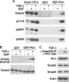
FIGURE 1.
FIGURE 1.
Pin1 binds to Smad2/3 in a TGF-β-dependent manner. A, TGF-β, but not EGF, induces Pin1-Smad3 interaction. HaCaT cells were treated with 300 pm TGF-β for 1 h or with 50 ng/ml EGF for 15 min for maximal induction of Smad3 phosphorylation in the linker region. The cell lysates were incubated with either GST or GST-Pin1 beads. The bound proteins were analyzed by immunoblot with an antibody against Smad3. The bound proteins were also analyzed by immunoblot with specific phosphopeptide antibodies against the phosphorylated Thr-179, Ser-204, and Ser-208 in the Smad3 linker region. B, TGF-β, but not EGF, induces Pin1-Smad2 interaction. HaCaT cells were treated with TGF-β or EGF as described in A. The cell lysates were incubated with GST or GST-Pin1 beads. The bound proteins were analyzed by immunoblot with an antibody against Smad2. The Smad2 levels in the cell lysates were also analyzed as a control. C, TGF-β induces Pin1-Smad2/3 interactions at endogenous protein levels. HaCaT cells were treated with or without TGF-β for 1 h. The cell lysates were immunoprecipitated with an antibody that was raised against the full-length Smad3 and recognizes both Smad3 and Smad2. The precipitates were analyzed by immunoblot with an antibody against Pin1. The expression levels of Pin1, Smad2, Smad3, and actin in the cell lysates were also analyzed as controls.
FIGURE 2.
FIGURE 2.
Pin1 binding to Smad3 requires both the linker and the C-tail phosphorylation, and the Thr-179 is the major binding site for Pin1. A, phosphorylation of both the linker and the C-tail in Smad3 is necessary for interaction with Pin1 in response to TGF-β. L17 cells were cotransfected with TβRI along with the wild type (WT) Smad3, a linker phosphorylation mutant (EPSM), or a C-tail phosphorylation mutant (C-tail 3A). Cells were then treated with or without 300 pm TGF-β for 1 h. The cell lysates were incubated with GST-Pin1 beads. The bound proteins were analyzed by immunoblot with the Smad3 antibody. B, Thr-179 is the major binding site in Smad3 for interaction with Pin1 in response to TGF-β. HEK293T cells were transfected with the wild type Smad3, a linker phosphorylation mutant Smad3, or a control plasmid that encodes GFP. Cells were treated with 300 pm TGF-β for 1 h. The cell lysates were incubated with the GST-Pin1 beads. The bound proteins were analyzed by immunoblot with the Smad3 antibody. The expression levels of the various Smad3 proteins were also analyzed as a control.
FIGURE 3.
FIGURE 3.
Knockdown of Pin1 does not have a significant effect on Smad2/3 levels, on TGF-β-induced linker and C-tail phosphorylation of Smad3, or on TGF-β-induced Smad2/3 nuclear accumulation. A, depletion of Pin1 has little effect on Smad2/3 levels in HaCaT cells. Stable HaCaT cell lines with an shRNA targeting Pin1 or the scrambled control were generated. The cell lysates were analyzed by immunoblot for Pin1, Smad2, and Smad3 levels. Actin levels were also analyzed as a control. B, depletion of Pin1 does not have a significant effect on TGF-β-induced linker and C-tail phosphorylation of Smad3 in HaCaT cells. The Pin1 knockdown HaCaT cells and the scrambled control HaCaT cells were treated with or without TGF-β for 1 h. The cell lysates were analyzed by immunoblot with phosphopeptide antibodies against the phosphorylated Thr-179, Ser-204, Ser-208, and the C-tail. Smad3 and Pin1 expression levels were also analyzed as controls. C, depletion of Pin1 does not affect TGF-β-induced Smad2/3 nuclear accumulation in HaCaT cells. The Pin1 knockdown HaCaT cells and the scrambled control HaCaT cells were treated with or without TGF-β for 1 h. Cells were then harvested and fractionated into the cytoplasmic (C) and nuclear (N) fractions. The same amount of proteins from the cytoplasmic fraction and nuclear fraction was analyzed for Smad2, Smad3, and Pin1 levels by immunoblot. GAPDH and proliferating cell nuclear antigen (PCNA) serve as cytoplasmic and nuclear markers, respectively.
FIGURE 4.
FIGURE 4.
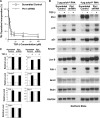
Knockdown of Pin1 does not have a significant effect on TGF-β-induced growth-inhibitory effects. A, knockdown of Pin1 does not have a significant effect on TGF-β-induced growth-inhibitory effects. Pin1 knockdown HaCaT cells and the scrambled control HaCaT cells were treated with or without TGF-β at various concentrations and then subjected to [3H]thymidine incorporation assay. The results represent the average of three independent experiments. B, knockdown of Pin1 does not have a significant effect on several TGF-β/Smad target genes. Pin1 knockdown HaCaT cells and the scrambled control HaCaT cells were treated with or without TGF-β for 8 h. The expression levels of Pin1 and several TGF-β/Smad target genes were analyzed by Northern blot analysis as indicated. GAPDH expression levels were also analyzed as a loading control. C, the mRNA levels of p15, p21, Smad7, JunB, PAI-1, Bcl-2, and Bub1 in B were quantified by densitometer and normalized to GAPDH mRNA levels. The bar graphs represent the average of results from 2 μg of poly(A)+ RNA and 4 μg of poly(A)+ RNA.
FIGURE 5.
FIGURE 5.
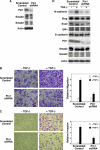
Knockdown of Pin1 significantly reduces TGF-β-induced migration and invasion. A, knockdown of Pin1 has little effect on Smad2/3 levels in the PC3 prostate cancer cells. Stable PC3 prostate cancer cell lines with the shRNA targeting Pin1 or the scrambled control were generated. The cell lysates were analyzed for expression levels of Pin1, Smad2, and Smad3. Actin levels were also analyzed as a loading control. B, knockdown of Pin1 significantly reduces TGF-β-induced migration of prostate cancer cells. The Pin1 knockdown PC3 cells and the scrambled control PC3 cells were subjected to the migration assay in the absence or presence of TGF-β. Representative photos of migrated cells were shown. The results from four independent experiments were plotted. C, knockdown of Pin1 significantly reduces TGF-β-induced invasion of prostate cancer cells. The Pin1 knockdown PC3 cells and the scrambled control PC3 cells were subjected to the invasion assay in the absence or presence of TGF-β. Representative photos of the invasion assay were shown. The results from four independent experiments were plotted. D, knockdown of Pin1 significantly reduces TGF-β induction of N-cadherin expression. The Pin1 knockdown PC3 cells and the scrambled control PC3 cells were treated with or without TGF-β. The cell lysates were analyzed by immunoblot with antibodies against N-cadherin, Slug, Snail, SIP1, E-cadherin, Pin1, Smad2, Smad3, and actin.
FIGURE 6.
FIGURE 6.
The catalytic activity of Pin1 is necessary for TGF-β-induced migration and invasion. A, the catalytic activity of Pin1 is necessary for TGF-β-induced migration. PC3 cells were subjected to the migration assay in the presence of 1 μm Pin1 inhibitor PiB or the vehicle DMSO control. Cells were treated with or without TGF-β. Representative photos of migrated cells were shown. The results from four independent experiments were plotted. B, catalytic activity of Pin1 is necessary for TGF-β-induced invasion. PC3 cells were subjected to the invasion assay in the presence of 1 μm PiB or the vehicle DMSO control. Cells were treated with or without TGF-β. Representative photos of the invasion assay are shown. The results from four independent experiments are plotted.

Similar articles

Cited by

References

    1. Roberts A. B., Sporn M. B. (1990) in Peptide Growth Factors and Their Receptors (Sporn M. B., Roberts A. B. eds) pp. 419–472, Springer-Verlag, Heidelberg, Germany
    1. Massagué J., Blain S. W., Lo R. S. (2000) Cell 103, 295–309 - PubMed
    1. Derynck R., Akhurst R. J., Balmain A. (2001) Nat. Genet. 29, 117–129 - PubMed
    1. Roberts A. B., Wakefield L. M. (2003) Proc. Natl. Acad. Sci. U.S.A. 100, 8621–8623 - PMC - PubMed
    1. Miyazono K., Suzuki H., Imamura T. (2003) Cancer Sci. 94, 230–234 - PMC - PubMed

Publication types

MeSH terms