. 2010 Feb 5;285(6):3973-3985.
doi: 10.1074/jbc.M109.064725. Epub 2009 Nov 17.

A conserved aromatic lock for the tryptophan rotameric switch in TM-VI of seven-transmembrane receptors

Affiliations

A conserved aromatic lock for the tryptophan rotameric switch in TM-VI of seven-transmembrane receptors

Birgitte Holst et al. J Biol Chem. .

Abstract

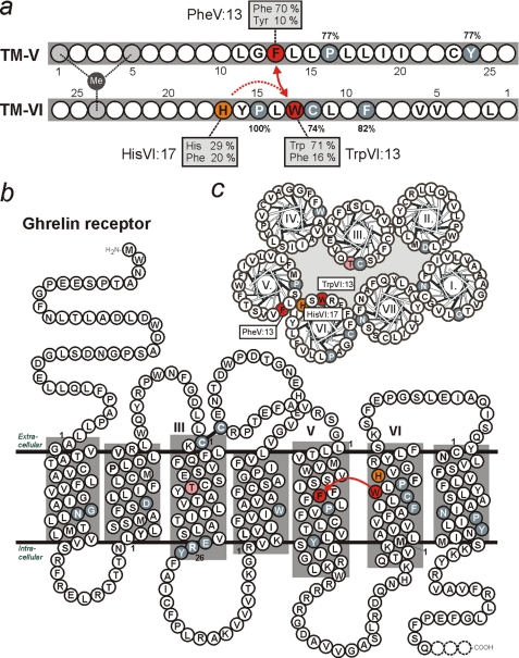
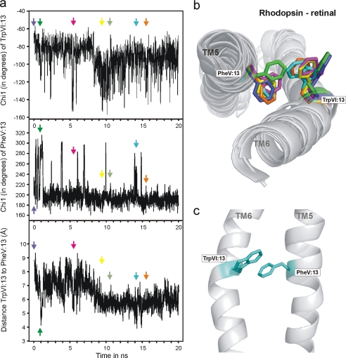
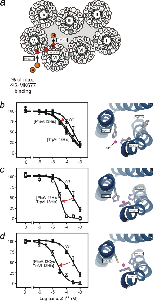
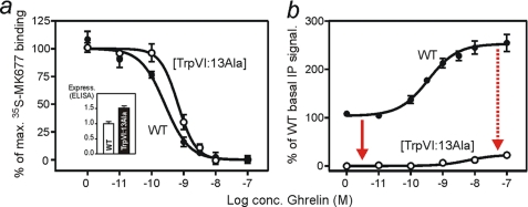
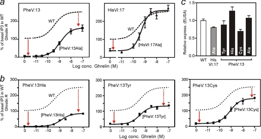
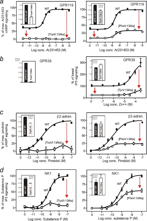
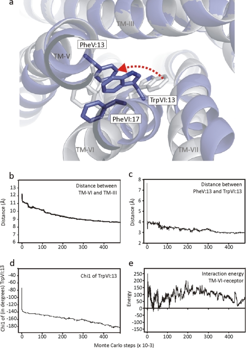
The conserved tryptophan in position 13 of TM-VI (Trp-VI:13 or Trp-6.48) of the CWXP motif located at the bottom of the main ligand-binding pocket in TM-VI is believed to function as a rotameric microswitch in the activation process of seven-transmembrane (7TM) receptors. Molecular dynamics simulations in rhodopsin demonstrated that rotation around the chi1 torsion angle of Trp-VI:13 brings its side chain close to the equally highly conserved Phe-V:13 (Phe-5.47) in TM-V. In the ghrelin receptor, engineering of high affinity metal-ion sites between these positions confirmed their close spatial proximity. Mutational analysis was performed in the ghrelin receptor with multiple substitutions and with Ala substitutions in GPR119, GPR39, and the beta(2)-adrenergic receptor as well as the NK1 receptor. In all of these cases, it was found that mutation of the Trp-VI:13 rotameric switch itself eliminated the constitutive signaling and strongly impaired agonist-induced signaling without affecting agonist affinity and potency. Ala substitution of Phe-V:13, the presumed interaction partner for Trp-VI:13, also in all cases impaired both the constitutive and the agonist-induced receptor signaling, but not to the same degree as observed in the constructs where Trp-VI:13 itself was mutated, but again without affecting agonist potency. In a proposed active receptor conformation generated by molecular simulations, where the extracellular segment of TM-VI is tilted inwards in the main ligand-binding pocket, Trp-VI:13 could rotate into a position where it obtained an ideal aromatic-aromatic interaction with Phe-V:13. It is concluded that Phe-V:13 can serve as an aromatic lock for the proposed active conformation of the Trp-VI:13 rotameric switch, being involved in the global movement of TM-V and TM-VI in 7TM receptor activation.


Figures

FIGURE 1.
FIGURE 1.
The location of the Trp-VI:13 rotameric switch and its putative interaction partner Phe-V:13 in the 7TM receptor structure. a, alignment of the amino acid sequence of oppositely oriented TM-V and TM-VI. In red is indicated Phe-V:13 (5.47), which is conserved as a Phe or Tyr residue in 80% of the 7TM family A receptors, and Trp-VI:13 (6.48), which is conserved as a Trp or Phe residue in 87% of the receptors. Position VI:176.52, which is occupied by a His (e.g. the ghrelin receptor) in 29% of the receptors and a Phe (e.g. the β2-adrenergic receptor) in 20% of the receptors is marked with orange. A few other highly conserved residues are highlighted in white on gray. The spatial proximity between residues in TM-V and TM-VI has previously been demonstrated by use of metal site engineering in positions V:01, V:04, and VI:24 (gray symbols) (33). b and c show serpentine and helical wheel diagrams of the ghrelin receptor, respectively. The residues indicated in white on gray represent the highly conserved “fingerprint residues” in each of the helices. The Phe-V:13 (5.47) and Trp-VI:13 (6.48) residues are highlighted in black on red, and the neighboring His-VI:17 is shown in black on orange. Thr-III:12, which is addressed under “Discussion” as an alternative interaction partner for the rotameric Trp-VI:13 switch, is highlighted in pink; no amino acid type is particularly conserved at this position except for Met (20%) (polar residues: Cys, 11%; Ser, 9%; Thr, 5%; Gln, 2%; Asn, 0%) (49).
FIGURE 2.
FIGURE 2.
Molecular dynamics simulations of the Trp-VI:13 rotameric switch and its interaction with Phe-V:13 in rhodopsin where 11-cis-retinal has been removed. a, chi1 torsion angle for Trp-VI:13, chi1 torsion angle for Phe-V:13, and distance between the indole group of Trp-VI:13 and the phenyl group of Phe-V:13, as measured from the center of the two aromatic rings, during 20 ns of molecular dynamics simulation of opsin. b, structures from various time points during the 20-ns molecular dynamics simulation showing the interaction between Trp-VI:13 and Phe-V:13. c, aromatic stacking of Trp-VI:13 and Phe-V:13 after 14-ns simulation.
FIGURE 3.
FIGURE 3.
Metal ion site engineering probing the proximity of positions V:13, VI:13, and VI:17 in the ghrelin receptor. a, helical wheel diagram, where the positions substituted by metal ion-chelating residues are indicated in black on red. b–d, competition binding experiments with Zn2+ (ZnCl2) performed in transiently transfected COS-7 cells using 35S-MK677 are shown for wild-type ghrelin receptor and the mutant forms of this with metal ion-chelating residues, His or Cys, substituted for Phe-V:13 and/or Trp-VI:13 as indicated. To the right in each of the panels is shown a molecular model of the putative metal ion-binding site; note that a metal ion-binding residue, His-VI:17, is found in the wild-type receptor background. The x-ray structure of the bovine rhodopsin was used as template for the molecular modeling.
FIGURE 4.
FIGURE 4.
Mutational analysis of Trp-VI:13 in the ghrelin receptor. a, competition binding experiment performed with 35S-MK677 as radioligand in COS-7 cells transiently transfected with either the wild-type (filled circles) or the Trp-VI:13 → Ala substituted ghrelin receptor (open circles). The inset shows cell surface expression of the receptors determined by ELISA under basal conditions. b, ghrelin-induced inositol phosphate accumulation in the wild-type ghrelin receptor and the Trp-VI:13 → Ala substituted receptor. The decrease in basal stimulation caused by Ala substitution of Trp-V:13 is indicated by a red arrow, and the decrease in ghrelin-induced maximum stimulation is indicated by a dotted red arrow.
FIGURE 5.
FIGURE 5.
Mutational analysis of Phe-V:13 and His-VI:17 in the ghrelin receptor. a, ghrelin-induced inositol phosphate accumulation of the wild-type ghrelin receptor (dashed line), the Phe-V:13 → Ala substituted receptor (black squares, left), and the His-VI:17 → Ala substituted receptor (black triangles, right) expressed as a percentage of the basal signaling in the wild-type receptor. b, ghrelin-induced inositol phosphate accumulation in three other mutant ghrelin receptors where Phe-V:13 was substituted with His (left), Tyr (middle), or Cys (right). In each of the panels, the decrease in basal stimulation caused by the mutations is indicated by a red arrow, and the decrease in ghrelin-induced maximum stimulation is indicated by a dotted red arrow. c, cell surface expression of mutant ghrelin receptors compared with wild-type receptor (open column) as measured by ELISA. All experiments were performed in transiently transfected COS-7 cells.
FIGURE 6.
FIGURE 6.
Mutational analysis of Trp-VI:13 and Phe-V:13 by Ala substitution in selected family A, rhodopsin-like 7TM receptors. a, the GPR119 receptor, which displays similar, high constitutive activity as the ghrelin receptor but signals through the Gαs pathway and is activated by lipid metabolites, and the non-peptide, small molecule agonist Ar231453 (55). The Ar231453-induced cAMP response is shown in the Trp-VI:13 → Ala (filled squares, left) and in the Phe-V:13 → Ala (filled circles, right) substituted receptors as compared with wild-type GPR119 receptor (open squares and open circles). b, the GPR39 receptor displays ∼25% constitutive signaling through the Gαq signaling pathway and is activated by Zn2+ (56). The Zn2+ induced inositol phosphate response is shown in the Phe-V:13 → Ala (filled circles, right) substituted receptor as compared with wild-type GPR39 receptor (open squares and open circles). The Trp- VI:13 → Ala mutant was poorly expressed (inset to the left), and its lack of response to Zn2+ is consequently not shown. c, the B2AR prototype family A 7TM receptor. The cAMP response in response to pindolol is shown in the Trp-VI:13 → Ala (filled squares, left) and in the Phe-V:13 → Ala (filled circles, right) substituted receptors as compared with wild-type B2AR (open symbols). d, the NK1 receptor, which displays undetectable constitutive activity, signals through the Gαq signaling pathway and is activated by substance P. The substance P-induced inositol phosphate response is shown in the Trp-VI:13 → Ala (filled squares, left) and in the Phe-V:13 → Ala (filled circles, right) substituted receptors as compared with the wild-type NK1 receptor (open squares and open circles). The insets in each panel show the cell surface expression relative to wild-type receptor as determined by ELISA. All experiments were performed in transiently transfected COS-7 cells.
FIGURE 7.
FIGURE 7.
Trp-VI:13 to Phe-V:13 interaction in a model of a presumed active conformation of the B2AR generated by Monte Carlo simulations. For the extracellular parts of the transmembrane segments, distance constraints corresponding to activating metal ion sites engineered between TM-III, TM-VI, and TM-VII were used, and for the intracellular parts, distance constraints based on the double electron-electron resonance measurements performed on double spin-labeled rhodopsin were used (30). As starting structure, we used the B2AR where Phe-VI:17 was manually rotated away from the binding pocket to its other preferred side chain conformation (see “Experimental Procedures”). a, in white ribbon is shown the starting structure, and in blue ribbon is shown the final conformation after the Monte Carlo simulation, and in both cases Phe-V:13, Trp-VI:13, and Phe-VI:17 are shown in stick models. The red dotted arrow indicates the rotation of Trp-VI:13 around its chi1 angle. b, the distance between the Cβ atoms of residue III:08 and VI:16, which constitute one of the distance constraints in the extracellular part of the receptor as determined. c, distance between the indole group of Trp-VI:13 and the phenyl group of Phe-V:13, as measured from the center of the two aromatic rings during the Monte Carlo simulation. d, the chi1 torsion angle of Trp-VI:13 determined throughout the Monte Carlo simulation. e, the interaction energy between TM-VI and the rest of the receptor, excluding internal energy contributions of TM-VI and the rest of the receptor calculated throughout the Monte Carlo simulation.

Similar articles

Cited by

References

    1. Schwartz T. W., Rosenkilde M. M. (1996) Trends Pharmacol. Sci. 17, 213–216 - PubMed
    1. Schwartz T. W., Frimurer T. M., Holst B., Rosenkilde M. M., Elling C. E. (2006) Annu. Rev. Pharmacol. Toxicol. 46, 481–519 - PubMed
    1. Pin J. P., Kniazeff J., Liu J., Binet V., Goudet C., Rondard P., Prézeau L. (2005) FEBS J. 272, 2947–2955 - PubMed
    1. Hubbell W. L., Altenbach C., Hubbell C. M., Khorana H. G. (2003) Adv. Protein Chem. 63, 243–290 - PubMed
    1. Kobilka B. K. (2007) Biochim. Biophys. Acta 1768, 794–807 - PMC - PubMed

Publication types

MeSH terms

LinkOut - more resources