. 2010 Jan 22;285(4):2847-56.
doi: 10.1074/jbc.M109.081141. Epub 2009 Nov 17.

Differential regulation of kainate receptor trafficking by phosphorylation of distinct sites on GluR6

Affiliations

Differential regulation of kainate receptor trafficking by phosphorylation of distinct sites on GluR6

Yukiko Nasu-Nishimura et al. J Biol Chem. .

Abstract

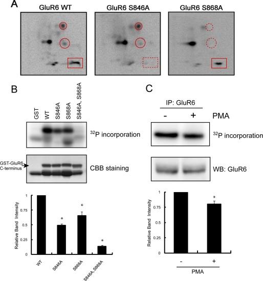
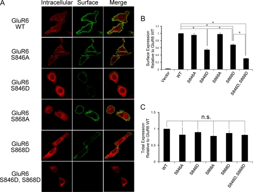
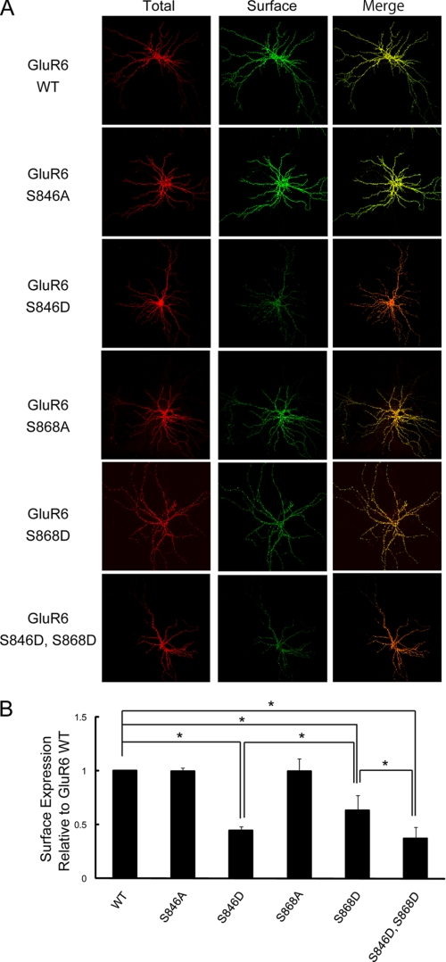
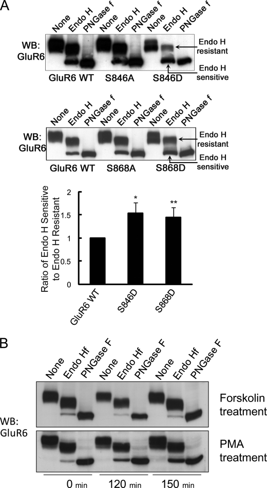
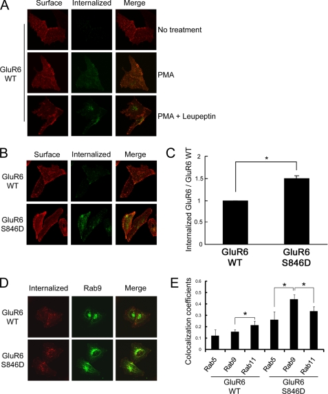
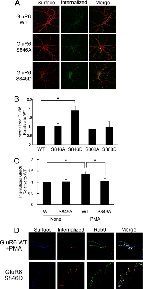
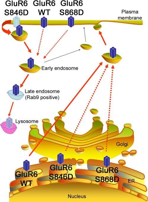
Kainate receptors are widely expressed in the brain, and are present at pre- and postsynaptic sites where they play a prominent role in synaptic plasticity and the regulation of network activity. Within individual neurons, kainate receptors of different subunit compositions are targeted to various locations where they serve distinct functional roles. Despite this complex targeting, relatively little is known about the molecular mechanisms regulating kainate receptor subunit trafficking. Here we investigate the role of phosphorylation in the trafficking of the GluR6 kainate receptor subunit. We identify two specific residues on the GluR6 C terminus, Ser(846) and Ser(868), which are phosphorylated by protein kinase C (PKC) and dramatically regulate GluR6 surface expression. By using GluR6 containing phosphomimetic and nonphosphorylatable mutations for these sites expressed in heterologous cells or in neurons lacking endogenous GluR6, we show that phosphorylation of Ser(846) or Ser(868) regulates receptor trafficking through the biosynthetic pathway. Additionally, Ser(846) phosphorylation dynamically regulates endocytosis of GluR6 at the plasma membrane. Our findings thus demonstrate that phosphorylation of PKC sites on GluR6 regulates surface expression of GluR6 at distinct intracellular trafficking pathways, providing potential molecular mechanisms for the PKC-dependent regulation of synaptic kainate receptor function observed during various forms of synaptic plasticity.


Figures

FIGURE 1.
FIGURE 1.
Identification of Ser846 and Ser868 as PKC phosphorylation sites in the GluR6 C terminus. A, phosphopeptide map analysis shows that the GluR6 C terminus is phosphorylated by PKC on multiple peptides (left panel) including Ser846 and Ser868, as indicated by the disappearance of phosphopeptides on maps of the GluR6 mutants (middle and right panels). GST-GluR6 (WT, S846A, and S868A) fusion proteins were phosphorylated with purified PKC in vitro using [γ-32P]ATP and then subjected to two-dimensional phosphopeptide mapping as described under “Experimental Procedures.” A rectangle surrounds the phosphopeptide that includes phosphorylated Ser846. Circles surround the phosphopeptides that include phosphorylated Ser868. An X in each panel indicates the origin where the peptides were initially spotted. Representative phosphopeptide maps are shown of 3–4 independent experiments. B, Ser846 and Ser868 are the major PKC phosphorylation sites in the GluR6 C terminus. GST, GST-GluR6 WT, GST-GluR6 S846A, GST-GluR6 S868A, and GST-GluR6 S846A,S868A were phosphorylated by PKC in vitro and analyzed by PhosphorImager. The loaded amount of GST fusion protein was visualized by protein staining. The lower band in the WT lane is a degradation product of GST-GluR6. Data represent mean ± S.E.M. (error bars) of normalized relative band intensity. *, p < 0.05, Student's t test. CCB, Coomassie Brilliant Blue. C, pretreatment of neurons with PMA reduced PKC phosphorylation of GluR6 in vitro. GluR6 was immunoprecipitated from PMA or vehicle-treated neurons, phosphorylated by PKC in vitro, and analyzed by PhosphorImager. The loaded amount of GluR6 was visualized by Western blotting using anti-GluR6 antibody. Data represent mean ± S.E.M. of normalized relative band intensity. *, p < 0.05, Student's t test.
FIGURE 2.
FIGURE 2.
GluR6 surface expression in HeLa cells was reduced when either Ser846 or Ser868 was mutated to mimic phosphorylation. A, GluR6 surface expression was evaluated in HeLa cells expressing FLAG-GluR6 (WT, S846A, S846D, S868A, S868D, or S846D,S868D) by immunofluorescence using confocal microscopy as described under “Experimental Procedures.” B, quantitative FACS analysis shows significantly decreased surface expression of GluR6 S846D and GluR6 S868D compared with GluR6 WT. HeLa cells were transfected with FLAG-GluR6 IRES-EGFP (GluR6 WT, S846A, S846D, S868A, S868D, or S846D,S868D). GluR6 surface expression was analyzed by FACS as described under “Experimental Procedures.” GluR6 S846D surface expression was decreased by 45%, GluR6 S868D by 31%, and GluR6 S846D,S868D by 69% compared with GluR6 WT. Data represent mean ± S.E.M. of the fold-increase in surface expression compared with GluR6 based on the fluorescence intensity of surface-expressed GluR6 in transfected cells. *, p < 0.01, Student's t test. C, quantitative FACS analysis shows that phosphomimetic mutations of GluR6 do not affect GluR6 total expression level. Data represent mean ± S.E.M. of the fold-increase in total expression compared with GluR6 WT based on the fluorescence intensity of total GluR6 in transfected cells.
FIGURE 3.
FIGURE 3.
GluR6 surface expression in neurons was reduced when either Ser846 or Ser868 was mutated to mimic phosphorylation. A, cultured hippocampal neurons derived from GluR6 KO mice were transfected with FLAG-GluR6 (WT, S846A, S846D, S868A, S868D, or S846D,S868D). Surface-expressed and total GluR6 were visualized by immunostaining as described under “Experimental Procedures.” B, quantification of surface-expressed GluR6 in GluR6 KO neurons using Metamorph software. The surface expression of GluR6 S846D, GluR6 S868D, and GluR6 S846D,S868D were significantly decreased compared with GluR6 WT (60% reduction for GluR6 S846D and 35% reduction for GluR6 S868D). Data represent mean ± S.E.M. of the fold-increase in surface expression compared with GluR6 WT based on the relative fluorescence intensity of surface-expressed GluR6 to that of total expressed GluR6. *, p < 0.01, Student's t test.
FIGURE 4.
FIGURE 4.
GluR6 S846D and GluR6 S868D are more highly retained in the ER than GluR6 WT. HeLa cells were transfected with FLAG-GluR6 (WT, S846A, S846D, S868A, or S868D). Cell lysates were prepared and subjected to a glycosidase assay as described under “Experimental Procedures.” Proteins were immunoblotted with GluR6 antibody. A, GluR6 S846D and GluR6 S868D were more endo H sensitive than GluR6 WT, GluR6 S846A, or GluR6 S868A. Endo H-sensitive fractions were quantitated by measuring the band intensity of the endo H-sensitive fraction compared with the band intensity of endo H-resistant fraction using NIH Image software. Error bars indicate S.E.M. *, p < 0.05, Student's t test. B, PKC activation increases ER retention of GluR6. Twenty-four hours after transfection, cells were treated with PMA (1 μm) and MG132 (20 μm) for the indicated time. GluR6 from PMA-treated cells was more endo H sensitive, as detected by a change in the ratio of immature GluR6 (lower band) to mature GluR6 (upper band) following endo H treatment. WB, Western blot.
FIGURE 5.
FIGURE 5.
Phosphorylation of Ser846 accelerates GluR6 internalization in HeLa cells. A, PMA treatment increases GluR6 internalization. GluR6 internalization was measured as described under “Experimental Procedures.” After surface labeling with anti-FLAG antibody, cells were incubated at 37 °C to allow labeled GluR6 to internalize for 60 min. Cells were fixed and incubated with fluorescence-conjugated secondary antibody for visualizing surface-expressed GluR6. After permeabilization, the cells were labeled with fluorescence-conjugated secondary antibody for visualizing internalized receptors. PMA treatment with leupeptin increased the accumulation of internalized GluR6. B, constitutive endocytosis of GluR6 S846D was more robust than GluR6 WT without PMA treatment. The brightness was adjusted equally across all panels in this figure to allow a direct comparison between GluR6 WT and GluR6 S846D. C, quantification of internalized GluR6 in HeLa cells was conducted using Metamorph software. GluR6 S846D showed a 1.5-fold increase in the normalized amount of internalized GluR6 compared with GluR6 WT. Error bars indicate S.E.M. *, p < 0.01. D, internalized GluR6 S846D was highly co-localized with a late endosomal marker, Rab9 in HeLa cells. HeLa cells were transfected with FLAG-GluR6 (WT or S846D) together with GFP-Rab9. Internalization assay of GluR6 (WT or S846D) in HeLa cells was performed following leupeptin treatment (60 min), and co-localization of internalized GluR6 with GFP-Rab9 was analyzed by immunocytochemistry. E, colocalization analysis was performed using LSM510 software (Zeiss). Values represent the mean ± S.E.M. of Pearson's correlation. *, p < 0.05.
FIGURE 6.
FIGURE 6.
Phosphorylation of Ser846 mediates GluR6 internalization in neurons. A, mutation of Ser846 to aspartate accelerates GluR6 internalization. Internalization of GluR6 (WT, S846A, and S846D) was evaluated in GluR6 KO neurons as described in the legend to Fig. 5A. The brightness was adjusted equally across all panels in this figure to allow direct comparison between GluR6 WT and S846D. B, quantification of internalized GluR6 was conducted using Metamorph software. GluR6 S846D showed an almost 2-fold increase in the normalized amount of internalized GluR6 compared with GluR6 WT. Error bars indicate S.E.M. *, p < 0.01. C, mutation of Ser846 to alanine blocks PMA-induced GluR6 internalization. Quantification of internalized GluR6 was conducted using Metamorph software. Error bars indicate S.E.M. *, p < 0.05. D, internalized GluR6 S846D was highly co-localized with the late endosomal marker Rab9 in neurons. Neurons were transfected with FLAG-GluR6 (WT or S846D) together with GFP-Rab9. The co-localization of internalized GluR6 WT with GFP-Rab9 was analyzed with immunofluorescence microscopy. Arrows indicate areas of colocalization.
FIGURE 7.
FIGURE 7.
Phosphorylation of GluR6 at Ser846 or Ser868 inhibits GluR6 exit from the ER and traffic to the cell surface. In addition, phosphorylation of GluR6 at Ser846 accelerates GluR6 endocytosis from the plasma membrane and traffic to late endosomes.

Similar articles

Cited by

References

    1. Lerma J., Paternain A. V., Rodríguez-Moreno A., López-García J. C. (2001) Physiol. Rev. 81, 971–998 - PubMed
    1. Vignes M., Collingridge G. L. (1997) Nature 388, 179–182 - PubMed
    1. Castillo P. E., Malenka R. C., Nicoll R. A. (1997) Nature 388, 182–186 - PubMed
    1. Chittajallu R., Vignes M., Dev K. K., Barnes J. M., Collingridge G. L., Henley J. M. (1996) Nature 379, 78–81 - PubMed
    1. Rodríguez-Moreno A., Herreras O., Lerma J. (1997) Neuron 19, 893–901 - PubMed

Publication types