. 2010 Jan 22;285(4):2397-414.
doi: 10.1074/jbc.M109.064295. Epub 2009 Nov 17.

Reciprocal negative regulation of PDK1 and ASK1 signaling by direct interaction and phosphorylation

Affiliations

Reciprocal negative regulation of PDK1 and ASK1 signaling by direct interaction and phosphorylation

Hyun-A Seong et al. J Biol Chem. .

Abstract

Cell survival and death-inducing signals are tightly associated with each other, and the decision as to whether a cell survives or dies is determined by controlling the relationship between these signals. However, the mechanism underlying the reciprocal regulation of such signals remains unclear. In this study, we reveal a functional association between PDK1 (3-phosphoinositide-dependent protein kinase 1), a critical mediator of cell survival, and ASK1 (apoptosis signal-regulating kinase 1), an apoptotic stress-activated MAPKKK. The physical association between PDK1 and ASK1 is mediated through the pleckstrin homology domain of PDK1 and the C-terminal regulatory domain of ASK1 and is decreased by ASK1-activating stimuli, such as H(2)O(2), tumor necrosis factor alpha, thapsigargin, and ionomycin, as well as insulin, a PDK1 stimulator. Wild-type PDK1, but not kinase-dead PDK1, negatively regulates ASK1 activity by phosphorylating Ser(967), a binding site for 14-3-3 protein, on ASK1. PDK1 functionally suppresses ASK1-mediated AP-1 transactivation and H(2)O(2)-mediated apoptosis in a kinase-dependent manner. On the other hand, ASK1 has been shown to inhibit PDK1 functions, including PDK1-mediated regulation of apoptosis and cell growth, by phosphorylating PDK1 at Ser(394) and Ser(398), indicating that these putative phosphorylation sites are involved in the negative regulation of PDK1 activity. These results provide evidence that PDK1 and ASK1 directly interact and phosphorylate each other and act as negative regulators of their respective kinases in resting cells.


Figures

FIGURE 1.
FIGURE 1.
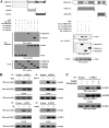
PDK1 forms a complex with ASK1 in vivo and in vitro. A, HEK293 cells were transiently transfected with the indicated combinations of expression vectors encoding GST-PDK1 and FLAG-ASK1. GST fusion proteins were purified with glutathione-Sepharose beads (GST Purification). Complex formation between PDK1 and ASK1 was determined by immunoblot analysis with an anti-FLAG antibody. B, proteins immunoprecipitated from HEK293 and HeLa cell lysates, with either rabbit preimmune serum (Preimm.) or anti-PDK1 antibody (α-PDK1), were immunoblotted with an anti-ASK1 antibody to determine the association between endogenous PDK1 and ASK1. C, for the in vitro association between PDK1 and ASK1, autophosphorylated recombinant PDK1 (2–3 μg) was incubated with unlabeled recombinant GST or GST-ASK1(K709R) (5 μg each) at room temperature for 1 h and analyzed on an 8% native polyacrylamide gel. WB, Western blot; IP, immunoprecipitation.
FIGURE 2.
FIGURE 2.
ASK1 and PDK1 stimuli destabilize the PDK1-ASK1 association mediated via their C-terminal domains. A, schematic representations of ASK1 and PDK1 divided into smaller HA- and FLAG-tagged fusion proteins, respectively. The numbers indicate amino acid residues corresponding to the domain boundaries. HEK293 cells were transiently transfected with the indicated combinations of expression vectors. Complex formation between PDK1 and ASK1 was determined by immunoblot analysis with an anti-HA antibody using GST precipitates (GST Purification) or FLAG immunoprecipitates (α-FLAG). B, HEK293 cell lysates were treated with or without the following stimuli: H2O2 (2 mm, 30 min), TNF-α (500 ng/ml, 30 min), thapsigargin (Tg; 20 μm, 30 min), or ionomycin (IONO; 1 μm, 24 h). C, HEK293 cells were incubated for 30 min with or without wortmannin (100 nm) and then treated with insulin (100 nm) for 20 min. The cell lysates were then immunoprecipitated with either rabbit preimmune serum (Preimm.) or anti-PDK1 antibody (α-PDK1), followed by immunoblotting with an anti-ASK1 antibody to determine the association between endogenous PDK1 and ASK1. WB, Western blot; IP, immunoprecipitation.
FIGURE 3.
FIGURE 3.
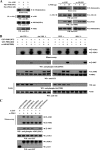
PDK1 inhibits ASK1 kinase activity. A, requirement of PDK1 kinase activity for PDK1-mediated regulation of ASK1 kinase activity. HEK293 cells were transiently transfected with HA-ASK1 in the presence or absence of Myc-tagged PDK1 (WT and KD). Cell lysates were then subjected to immunoprecipitation with an anti-HA antibody, and the resulting immunoprecipitates were subjected to an in vitro kinase assay using GST-tagged MKK6(K82A) as the substrate to determine ASK1 kinase activity (left). Approximately 5 μg of recombinant GST-tagged wild-type ASK1 (re.ASK1) or ASK1(K) (re.ASK1(K)) proteins were incubated at room temperature for 1 h with the amount of recombinant GST-tagged PDK1 (re.PDK1) indicated in 30 μl of 20 mm Tris-HCl buffer (pH 7.5), and ASK1 kinase activity was determined by an in vitro kinase assay using GST-tagged MKK6(K82A) as a substrate (right). The circled letters P indicate phosphorylation. B, PDK1 phosphorylates ASK1. HEK293 cells transiently expressing GST-tagged PDK1 (WT and KD) were treated (+) or untreated (−) with specific Akt inhibitors (VIII, 20 μm; SH-5, 10 μm) for 30 min. GST-PDK1 (WT and KD), precipitated using glutathione-Sepharose beads, was assayed for its kinase activity in the presence of kinase buffer containing ∼3–4 μg of recombinant GST-tagged ASK1(K709R) substrate. C, identification of PDK1 phosphorylation sites on ASK1. The recombinant GST-tagged PDK1 (re.PDK1) was analyzed for its kinase activity by an in vitro kinase assay using recombinant GST-tagged ASK1(K709R) and one of its substitution mutants, ASK1(S83A), ASK1(T838A), ASK1(S967A), or ASK1(S1034A), as substrates. GST alone was used as a nonspecific control. WB, Western blot; IP, immunoprecipitation.
FIGURE 4.
FIGURE 4.
PDK1 inhibits ASK1 downstream signaling. Transiently transfected HEK293 cells expressing an empty vector (Vector), PDK1(WT), or PDK1(KD) (A); parental HEK293 cells (B and C); and HEK293 cells (PDK1(OE)) stably overexpressing PDK1 or HEK293 cells (PDK1 shRNA) stably expressing PDK1-specific shRNA (B and C) were incubated with or without 2 mm H2O2 for 30 min. The activities of ASK1, MKK3, MKK6, or p38 were determined by in vitro kinase assays or immunoblot analyses using the immunoprecipitates indicated or total cell lysates treated (+) or untreated (−) with a specific Akt inhibitor VIII (Akt VIII). The overexpression or knockdown level of endogenous PDK1 was determined by anti-PDK1 immunoblotting (see supplemental Fig. 3). The relative level of kinase activity was quantitated by densitometric analyses, and the -fold increase relative to untreated samples in control cells containing an empty vector alone or parental HEK293 cells was calculated. The circled letters P indicate phosphorylation. WB, Western blot; IP, immunoprecipitation.
FIGURE 4.
FIGURE 4.
PDK1 inhibits ASK1 downstream signaling. Transiently transfected HEK293 cells expressing an empty vector (Vector), PDK1(WT), or PDK1(KD) (A); parental HEK293 cells (B and C); and HEK293 cells (PDK1(OE)) stably overexpressing PDK1 or HEK293 cells (PDK1 shRNA) stably expressing PDK1-specific shRNA (B and C) were incubated with or without 2 mm H2O2 for 30 min. The activities of ASK1, MKK3, MKK6, or p38 were determined by in vitro kinase assays or immunoblot analyses using the immunoprecipitates indicated or total cell lysates treated (+) or untreated (−) with a specific Akt inhibitor VIII (Akt VIII). The overexpression or knockdown level of endogenous PDK1 was determined by anti-PDK1 immunoblotting (see supplemental Fig. 3). The relative level of kinase activity was quantitated by densitometric analyses, and the -fold increase relative to untreated samples in control cells containing an empty vector alone or parental HEK293 cells was calculated. The circled letters P indicate phosphorylation. WB, Western blot; IP, immunoprecipitation.
FIGURE 5.
FIGURE 5.
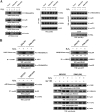
PDK1-mediated inhibition of ASK1 activity is dependent on the modulation of the association between ASK1 and its regulators, 14-3-3 and MKK3, and ASK1 phosphorylation. A and B, modulation of the endogenous association of ASK1 with 14-3-3 protein and MKK3. HEK293 cells were transfected with the indicated combinations of FLAG-14-3-3, FLAG-ASK1, HA-MKK3, HA-ASK1, Myc-tagged PDK1 (WT and KD), GST-PDK1, and PDK1-specific siRNA. Cell lysates were then subjected to immunoprecipitation with the antibodies indicated, and the resulting immunoprecipitates were analyzed by immunoblot analysis using the appropriate antibodies to determine the association of 14-3-3 protein (A) and MKK3 (B) with ASK1. The knockdown effect of endogenous PDK1 on the association between endogenous ASK1 and 14-3-3 protein or MKK3 was determined by immunoblot analysis with the antibodies indicated using HEK293 cells transiently transfected with PDK1-specific siRNA 1 (A and B, right panels). C, HEK293 cells were transiently transfected with the following expression vectors: empty vector (Vector), PDK1 (WT and KD), or PDK1 siRNAs (#1 and #2, each 200 nm) in the presence of ASK1. The cells were then treated with or without 2 mm H2O2 for 30 min. ASK1 phosphorylation was determined by immunoblot analysis using anti-phospho-ASK1(Ser83), anti-phospho-ASK1(Thr845), and anti-phospho-ASK1(Ser967) antibodies. D, parental HEK293 cells or HEK293 cells (PDK1 shRNA) stably expressing PDK1-specific shRNA were incubated for 30 min in the presence or absence of a specific Akt inhibitor VIII (20 μm). Cell lysates were examined for ASK1 phosphorylation (Ser83, Thr838, and Ser967) by immunoblot analysis using the antibodies indicated. The knockdown level of endogenous PDK1 was determined by anti-PDK1 immunoblotting (fourth panel). The relative level of phosphorylation was quantitated by densitometric analyses, and the -fold increase relative to untreated control cells expressing an empty vector alone in the presence of ASK1 or parental cells was calculated. The circled letters P indicate phosphorylation. WB, Western blot; IP, immunoprecipitation.
FIGURE 5.
FIGURE 5.
PDK1-mediated inhibition of ASK1 activity is dependent on the modulation of the association between ASK1 and its regulators, 14-3-3 and MKK3, and ASK1 phosphorylation. A and B, modulation of the endogenous association of ASK1 with 14-3-3 protein and MKK3. HEK293 cells were transfected with the indicated combinations of FLAG-14-3-3, FLAG-ASK1, HA-MKK3, HA-ASK1, Myc-tagged PDK1 (WT and KD), GST-PDK1, and PDK1-specific siRNA. Cell lysates were then subjected to immunoprecipitation with the antibodies indicated, and the resulting immunoprecipitates were analyzed by immunoblot analysis using the appropriate antibodies to determine the association of 14-3-3 protein (A) and MKK3 (B) with ASK1. The knockdown effect of endogenous PDK1 on the association between endogenous ASK1 and 14-3-3 protein or MKK3 was determined by immunoblot analysis with the antibodies indicated using HEK293 cells transiently transfected with PDK1-specific siRNA 1 (A and B, right panels). C, HEK293 cells were transiently transfected with the following expression vectors: empty vector (Vector), PDK1 (WT and KD), or PDK1 siRNAs (#1 and #2, each 200 nm) in the presence of ASK1. The cells were then treated with or without 2 mm H2O2 for 30 min. ASK1 phosphorylation was determined by immunoblot analysis using anti-phospho-ASK1(Ser83), anti-phospho-ASK1(Thr845), and anti-phospho-ASK1(Ser967) antibodies. D, parental HEK293 cells or HEK293 cells (PDK1 shRNA) stably expressing PDK1-specific shRNA were incubated for 30 min in the presence or absence of a specific Akt inhibitor VIII (20 μm). Cell lysates were examined for ASK1 phosphorylation (Ser83, Thr838, and Ser967) by immunoblot analysis using the antibodies indicated. The knockdown level of endogenous PDK1 was determined by anti-PDK1 immunoblotting (fourth panel). The relative level of phosphorylation was quantitated by densitometric analyses, and the -fold increase relative to untreated control cells expressing an empty vector alone in the presence of ASK1 or parental cells was calculated. The circled letters P indicate phosphorylation. WB, Western blot; IP, immunoprecipitation.
FIGURE 6.
FIGURE 6.
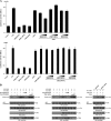
PDK1 suppresses JNK-mediated transcription and H2O2-induced apoptosis. A, 293T cells were transiently transfected with the indicated combinations of expression vectors encoding c-fos (0.6 μg), PDK1 (WT and KD) (0.2 and 0.6 μg), wild-type and mutant (S967A) ASK1 (each 0.6 μg), and PDK1-specific siRNA 1 (50 and 200 nm) together with AP-1 luciferase plasmid (0.2 μg) and an expression plasmid (0.2 μg) for β-galactosidase as an internal control. Luciferase activity was measured 48 h after transfection and normalized to β-galactosidase activity. B, inhibition of JNK activity by PDK1. HEK293 cells were transfected with HA-ASK1 and GST-JNK in the presence or absence of increasing amounts of PDK1 (WT and KD) or PDK1-specific siRNA 1. Cell lysates were prepared and analyzed for JNK activity by an in vitro kinase assay using c-Jun as a substrate. JNK activity was also determined in PDK1 knockdown cells (PDK1 shRNA) transfected with HA-ASK1 and GST-JNK in the presence or absence of H2O2 (right). C and D, HEK293 cells transiently expressing the indicated combinations of PDK1 (WT and KD) (1 and 3 μg), wild-type ASK1 (1 and 3 μg), and PDK1- and ASK1-specific siRNAs (100 and 200 nm) were incubated with or without H2O2 (1 mm) for 9 h in the presence or absence of a specific Akt inhibitor VIII (20 μm). Apoptotic cell death was determined using the GFP expression system (GFP) or terminal deoxynucleotide transferase-mediated dUTP nick end labeling (TUNEL), as described under “Materials and Methods.” E, HEK293 cells harboring stably integrated pSingle-tTS-shRNA empty vector (Vector) or pSingle-tTS-shRNA vector containing PDK1-specific shRNA (PDK1 shRNA (inducible)) were cultured in the presence or absence of 1 μg/ml doxycycline (Dox) for 72 h to determine the activities of ASK1, MKK3, MKK6, or p38. Inducible silencing of endogenous PDK1 expression by doxycycline was assessed by immunoblotting using an anti-PDK1 antibody. β-Actin was used as a loading control. The circled letters P indicate phosphorylation. WB, Western blot; RLU, relative luciferase units.
FIGURE 6.
FIGURE 6.
PDK1 suppresses JNK-mediated transcription and H2O2-induced apoptosis. A, 293T cells were transiently transfected with the indicated combinations of expression vectors encoding c-fos (0.6 μg), PDK1 (WT and KD) (0.2 and 0.6 μg), wild-type and mutant (S967A) ASK1 (each 0.6 μg), and PDK1-specific siRNA 1 (50 and 200 nm) together with AP-1 luciferase plasmid (0.2 μg) and an expression plasmid (0.2 μg) for β-galactosidase as an internal control. Luciferase activity was measured 48 h after transfection and normalized to β-galactosidase activity. B, inhibition of JNK activity by PDK1. HEK293 cells were transfected with HA-ASK1 and GST-JNK in the presence or absence of increasing amounts of PDK1 (WT and KD) or PDK1-specific siRNA 1. Cell lysates were prepared and analyzed for JNK activity by an in vitro kinase assay using c-Jun as a substrate. JNK activity was also determined in PDK1 knockdown cells (PDK1 shRNA) transfected with HA-ASK1 and GST-JNK in the presence or absence of H2O2 (right). C and D, HEK293 cells transiently expressing the indicated combinations of PDK1 (WT and KD) (1 and 3 μg), wild-type ASK1 (1 and 3 μg), and PDK1- and ASK1-specific siRNAs (100 and 200 nm) were incubated with or without H2O2 (1 mm) for 9 h in the presence or absence of a specific Akt inhibitor VIII (20 μm). Apoptotic cell death was determined using the GFP expression system (GFP) or terminal deoxynucleotide transferase-mediated dUTP nick end labeling (TUNEL), as described under “Materials and Methods.” E, HEK293 cells harboring stably integrated pSingle-tTS-shRNA empty vector (Vector) or pSingle-tTS-shRNA vector containing PDK1-specific shRNA (PDK1 shRNA (inducible)) were cultured in the presence or absence of 1 μg/ml doxycycline (Dox) for 72 h to determine the activities of ASK1, MKK3, MKK6, or p38. Inducible silencing of endogenous PDK1 expression by doxycycline was assessed by immunoblotting using an anti-PDK1 antibody. β-Actin was used as a loading control. The circled letters P indicate phosphorylation. WB, Western blot; RLU, relative luciferase units.
FIGURE 7.
FIGURE 7.
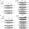
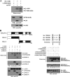
ASK1 phosphorylates PDK1. A, for the in vitro kinase assay, ∼5 μg of recombinant PDK1(KD) proteins were mixed with 20 μm ATP, 0.3 μCi of [γ-32P]ATP, and 20 mm MgCl2 in 30 μl of kinase buffer and incubated with the HA immunoprecipitates for HA-ASK1 at 30 °C for 30 min with frequent gentle mixing. B, identification of ASK1 phosphorylation sites on PDK1. The recombinant ASK1 protein was analyzed for its kinase activity by an in vitro kinase assay using recombinant PDK1-N, PDK1-C, substitution mutants (S258A/S262A, S394A/S398A, or T518A/T522A) of PDK1-C, and the positive control MKK6(K82A) as substrates. C, effect of ASK1 on PI3K/PDK1-mediated signaling. HEK293 cells, transfected with the expression vectors indicated, were lysed, and the GST precipitates were then analyzed for PDK1 activity by an in vitro kinase assay using serum/glucocorticoid-regulated kinase as a substrate and by immunoblot analysis with an anti-phospho-PDK1(Ser241) antibody (left). The Akt and Bad immunoprecipitates were also analyzed for Akt and Bad activation by immunoblotting with anti-phospho-Akt(Thr308) and anti-phospho-Bad(Ser136) antibodies (middle and right). The circled letters P indicate phosphorylation. re., recombinant. WB, Western blot.
FIGURE 7.
FIGURE 7.
ASK1 phosphorylates PDK1. A, for the in vitro kinase assay, ∼5 μg of recombinant PDK1(KD) proteins were mixed with 20 μm ATP, 0.3 μCi of [γ-32P]ATP, and 20 mm MgCl2 in 30 μl of kinase buffer and incubated with the HA immunoprecipitates for HA-ASK1 at 30 °C for 30 min with frequent gentle mixing. B, identification of ASK1 phosphorylation sites on PDK1. The recombinant ASK1 protein was analyzed for its kinase activity by an in vitro kinase assay using recombinant PDK1-N, PDK1-C, substitution mutants (S258A/S262A, S394A/S398A, or T518A/T522A) of PDK1-C, and the positive control MKK6(K82A) as substrates. C, effect of ASK1 on PI3K/PDK1-mediated signaling. HEK293 cells, transfected with the expression vectors indicated, were lysed, and the GST precipitates were then analyzed for PDK1 activity by an in vitro kinase assay using serum/glucocorticoid-regulated kinase as a substrate and by immunoblot analysis with an anti-phospho-PDK1(Ser241) antibody (left). The Akt and Bad immunoprecipitates were also analyzed for Akt and Bad activation by immunoblotting with anti-phospho-Akt(Thr308) and anti-phospho-Bad(Ser136) antibodies (middle and right). The circled letters P indicate phosphorylation. re., recombinant. WB, Western blot.
FIGURE 8.
FIGURE 8.
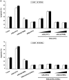
ASK1 negatively regulates PDK1-dependent apoptosis and cell growth. A, effect of ASK1 on PDK1-mediated apoptosis. 293T cells were transiently transfected with the expression vectors indicated. After 24 h, cells were treated with TNF-α (20 ng/ml) and cycloheximide (10 μg/ml) for 14 h to induce apoptosis. Apoptotic cell death was determined by the GFP system and TUNEL staining. B, effect of ASK1 on PDK1-mediated cell cycle progression. HaCaT cells (2 × 10⁵/dish), transfected with the indicated combinations of plasmid vectors, were synchronized in G0/G1 by hydroxyurea treatment (30). Cells were collected after 10% serum treatment for 24 h and evaluated for determination of cell numbers in G0/G1, S, and G2/M phases by flow cytometry analysis. PDK1(S394A/S398A), the wild-type PDK1 mutant S394A/S398A. C, ASK1-intact MEFs or ASK1-deficient MEFs were incubated for 30 min with or without 100 nm wortmannin and then treated with 100 nm insulin for 20 min. The cell lysates were subjected to immunoblot analysis with the antibodies indicated. β-Actin was used as a loading control. D, a model for the regulation of PDK1-ASK1 complex formation in response to signals that act on PDK1 or ASK1. The dotted and solid lines represent ASK1 and PDK1 signals, respectively. Details of this model are given under “Discussion.” WB, Western blot.
FIGURE 8.
FIGURE 8.
ASK1 negatively regulates PDK1-dependent apoptosis and cell growth. A, effect of ASK1 on PDK1-mediated apoptosis. 293T cells were transiently transfected with the expression vectors indicated. After 24 h, cells were treated with TNF-α (20 ng/ml) and cycloheximide (10 μg/ml) for 14 h to induce apoptosis. Apoptotic cell death was determined by the GFP system and TUNEL staining. B, effect of ASK1 on PDK1-mediated cell cycle progression. HaCaT cells (2 × 10⁵/dish), transfected with the indicated combinations of plasmid vectors, were synchronized in G0/G1 by hydroxyurea treatment (30). Cells were collected after 10% serum treatment for 24 h and evaluated for determination of cell numbers in G0/G1, S, and G2/M phases by flow cytometry analysis. PDK1(S394A/S398A), the wild-type PDK1 mutant S394A/S398A. C, ASK1-intact MEFs or ASK1-deficient MEFs were incubated for 30 min with or without 100 nm wortmannin and then treated with 100 nm insulin for 20 min. The cell lysates were subjected to immunoblot analysis with the antibodies indicated. β-Actin was used as a loading control. D, a model for the regulation of PDK1-ASK1 complex formation in response to signals that act on PDK1 or ASK1. The dotted and solid lines represent ASK1 and PDK1 signals, respectively. Details of this model are given under “Discussion.” WB, Western blot.

Similar articles

Cited by

References

    1. Brunet A., Bonni A., Zigmond M. J., Lin M. Z., Juo P., Hu L. S., Anderson M. J., Arden K. C., Blenis J., Greenberg M. E. (1999) Cell 96, 857–868 - PubMed
    1. Cardone M. H., Roy N., Stennicke H. R., Salvesen G. S., Franke T. F., Stanbridge E., Frisch S., Reed J. C. (1998) Science 282, 1318–1321 - PubMed
    1. Cross D. A., Alessi D. R., Cohen P., Andjelkovich M., Hemmings B. A. (1995) Nature 378, 785–789 - PubMed
    1. Datta S. R., Dudek H., Tao X., Masters S., Fu H., Gotoh Y., Greenberg M. E. (1997) Cell 91, 231–241 - PubMed
    1. Hetman M., Cavanaugh J. E., Kimelman D., Xia Z. (2000) J. Neurosci. 20, 2567–2574 - PMC - PubMed

Publication types

MeSH terms