. 2010 Jan 15;285(3):1939-49.
doi: 10.1074/jbc.M109.057216. Epub 2009 Nov 17.

The amyloid precursor protein/protease nexin 2 Kunitz inhibitor domain is a highly specific substrate of mesotrypsin

Affiliations

The amyloid precursor protein/protease nexin 2 Kunitz inhibitor domain is a highly specific substrate of mesotrypsin

Moh'd A Salameh et al. J Biol Chem. .

Abstract

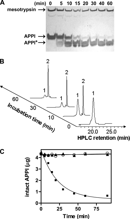
The amyloid precursor protein (APP) is a ubiquitously expressed transmembrane adhesion protein and the progenitor of amyloid-beta peptides. The major splice isoforms of APP expressed by most tissues contain a Kunitz protease inhibitor domain; secreted APP containing this domain is also known as protease nexin 2 and potently inhibits serine proteases, including trypsin and coagulation factors. The atypical human trypsin isoform mesotrypsin is resistant to inhibition by most protein protease inhibitors and cleaves some inhibitors at a substantially accelerated rate. Here, in a proteomic screen to identify potential physiological substrates of mesotrypsin, we find that APP/protease nexin 2 is selectively cleaved by mesotrypsin within the Kunitz protease inhibitor domain. In studies employing the recombinant Kunitz domain of APP (APPI), we show that mesotrypsin cleaves selectively at the Arg(15)-Ala(16) reactive site bond, with kinetic constants approaching those of other proteases toward highly specific protein substrates. Finally, we show that cleavage of APPI compromises its inhibition of other serine proteases, including cationic trypsin and factor XIa, by 2 orders of magnitude. Because APP/protease nexin 2 and mesotrypsin are coexpressed in a number of tissues, we suggest that processing by mesotrypsin may ablate the protease inhibitory function of APP/protease nexin 2 in vivo and may also modulate other activities of APP/protease nexin 2 that involve the Kunitz domain.


Figures

FIGURE 1.
FIGURE 1.
Identification of APP as a mesotrypsin substrate. A, conditioned medium from NB11 prostate cancer cells was enriched for trypsin-binding proteins by affinity chromatography as described under “Experimental Procedures.” Resultant protein samples, either untreated (left lane) or subjected to digestion with 50 nm mesotrypsin at 37 °C for 1 h (right lane), were resolved by SDS-PAGE and silver-stained, and bands differing between the two lanes were identified by nanoLC-MS/MS as described under “Experimental Procedures.” The band labeled APP was identified with 33 unique peptide matches to the human APP sequence; bands labeled B40 and B65 were tentatively identified as N-terminal and C-terminal proteolytic fragments derived from APP. B, the complete sequence of the human APP gene, showing coverage of peptides identified by nanoLC-MS/MS in boldface type and underlined. The Kunitz domain sequence is shown in red, with the reactive site Arg residue capitalized.
FIGURE 2.
FIGURE 2.
Hydrolysis of sAPP by mesotrypsin. Conditioned media from prostate cancer cell lines were treated with mesotrypsin and analyzed for APP protein on Western blots. Serum-free conditioned media from confluent WPE1-NB11 cells (A) or LNCaP cells (B) were collected after 5 days, concentrated 6-fold, and treated for 1 h at 37 °C with recombinant mesotrypsin at the concentrations shown. Western blots probed with antibody 22C11, which recognizes an N-terminal APP epitope, show a mesotrypsin-dependent disappearance of intact sAPP (∼120 kDa), along with concomitant accumulation of a cleavage fragment of ∼40 kDa. C, incubation of WPE1-NB11 conditioned medium was performed as described in A with the following recombinant enzymes: buffer only (lane 1), mesotrypsin inhibited with 10 mm phenylmethylsulfonyl fluoride (lane 2), mesotrypsin-S195A (lane 3), cationic trypsin (lane 4), and mesotrypsin (lane 5). All enzymes were at 100 nm concentration.
FIGURE 3.
FIGURE 3.
Hydrolysis of recombinant APPI by mesotrypsin. A, SDS-polyacrylamide gel shows a time course of APPI hydrolysis by mesotrypsin. APPI (25 μm) and mesotrypsin (250 nm) were incubated at 37 °C, and then samples were withdrawn and quenched at various time points for subsequent analysis. The arrows indicate mesotrypsin (24 kDa), APPI (6.3 kDa), and APPI* hydrolysis products. B, representative HPLC chromatograms are shown from a similar time course of APPI hydrolysis by mesotrypsin. Peak 1 eluting at 21.5 min and peak 2 eluting at 23.0 min were isolated and subjected to mass spectrometry and N-terminal sequencing to confirm that they represent intact APPI and cleaved APPI*, respectively (see Fig. 4 and Table 1). C, disappearance of intact APPI was quantified by integration of HPLC peak 1 in a time course similar to that illustrated in B, in incubations of APPI with mesotrypsin (■), the catalytically inactive mesotrypsin-S195A (○), or cationic trypsin (▴). Equilibrium was attained between intact and cleaved APPI over the course of the 90-min incubation with mesotrypsin. APPI concentration was 25 μm, and enzyme concentration was 250 nm in each incubation.
FIGURE 4.
FIGURE 4.
Scheme for identification of Arg15-Ala16 as the mesotrypsin cleavage site within APPI. The complete sequence of APPI is shown, with intramolecular disulfide bonds indicated by black brackets below the sequence. The Arg15-Ala16 reactive site bond, anticipated to be specifically targeted for cleavage by mesotrypsin, is indicated by the black arrow. As illustrated by the scheme, mesotrypsin cleavage yields a single species composed of two peptide chains covalently linked by two disulfide bonds; because a water molecule has been incorporated in the enzymatic hydrolysis reaction, this APPI* species is 18 Da higher in mass than the APPI precursor. Following reduction of the disulfides, the peptide chains can be isolated by HPLC and individually characterized. The depicted species APPI (HPLC peak 1 from Fig. 3B), APPI* (HPLC peak 2 from Fig. 3B), and the C-terminal fragment were isolated and analyzed for intact mass and N-terminal sequence, with the results shown in Table 1.
FIGURE 5.
FIGURE 5.
Kinetics of APPI inhibition of and hydrolysis by mesotrypsin. A, mesotrypsin cleavage of peptide substrate Z-GPR-pNA is competitively inhibited by APPI with a Ki of 136 nm; because APPI is an alternative, competing substrate for mesotrypsin, this value also represents the Km for mesotrypsin cleavage of APPI. APPI concentration was 0 nm (□), 50 nm (▾), 100 nm (○), 200 nm (■), or 400 nm (▵); mesotrypsin concentration was 0.25 nm. Data were fit globally to the competitive inhibition equation; the Lineweaver-Burk double reciprocal transform is shown here. B, mesotrypsin cleavage of Z-GPR-pNA is only minimally inhibited by 1 mm Gly-Pro-Ser-Arg-Ala-Met-Ile-Tyr, a peptide mimic of the APPI binding loop. Peptide concentration was 0 mm (▵) or 1 mm (■); mesotrypsin concentration was 0.25 nm. C, disappearance of intact APPI in a time course similar to that illustrated in Fig. 3B was quantified by HPLC peak integration. The linear initial rate of hydrolysis, observed under conditions of enzyme saturation, allows calculation of a catalytic rate constant kcat of 4.2 × 10⁻² s−1. APPI and mesotrypsin concentrations were 25 μm and 25 nm, respectively. D, disappearance of intact APPI in a time course incubation with cationic trypsin was quantified by HPLC peak integration. The linear initial rate of hydrolysis, observed under conditions of enzyme saturation, allows calculation of a catalytic rate constant kcat of 1.8 × 10⁻⁵ s−1. APPI and cationic trypsin concentrations were 50 μm and 5 μm, respectively.
FIGURE 6.
FIGURE 6.
APPI* cleaved at Arg15-Ala16 is severely compromised as an inhibitor of cationic trypsin. A and B, intact APPI inhibited cationic trypsin as a slow, tight binding inhibitor with a Ki of 170 pm. A, steady-state equilibrium rates were obtained from reactions including various concentrations of APPI as noted on the plot; reactions included 0.1 nm cationic trypsin and 150 μm Z-GPR-pNA substrate. B, The replot of (v0vi)/vi versus [APPI], where v0 is the uninhibited rate and vi is the rate in the presence of APPI, allows calculation of Ki as described under “Experimental Procedures.” C, cleaved APPI* is a classic competitive inhibitor of cationic trypsin, with an inhibition constant Ki of 22 nm. APPI* concentration was 0 nm (□), 10 nm (▾), 25 nm (○), or 50 nm (■); cationic trypsin concentration was 0.8 nm. Data were fitted as in Fig. 5A.
FIGURE 7.
FIGURE 7.
APPI* cleaved at Arg15-Ala16 is severely compromised as an inhibitor of factor XIa. Equilibrium binding constants were determined by preincubating FXIa (25 pm) with increasing concentrations of intact APPI (A) or cleaved APPI* (B) for 30 min at 37 °C and then measuring the remaining FXIa activity toward t-butoxycarbonyl-Glu(benzyl ester)-Ala-Arg-4-methylcoumaryl-7-amide (275 μm). Points represent mean values ± S.D. of four assays each. C, effects of APPI and APPI* on the clotting time in an APTT assay are compared. Values are reported as -fold increase compared with controls in the absence of inhibitors; points represent mean values ± S.D. of four determinations for APPI (solid circles) and five determinations for APPI* (solid squares). Assays were carried out in microcuvettes at 37 °C using normal pooled plasma.

Similar articles

Cited by

References

    1. Chen J. M., Ferec C. (2000) Pancreas 21, 57–62 - PubMed
    1. Ge X., Yamamoto S., Tsutsumi S., Midorikawa Y., Ihara S., Wang S. M., Aburatani H. (2005) Genomics 86, 127–141 - PubMed
    1. Su A. I., Wiltshire T., Batalov S., Lapp H., Ching K. A., Block D., Zhang J., Soden R., Hayakawa M., Kreiman G., Cooke M. P., Walker J. R., Hogenesch J. B. (2004) Proc. Natl. Acad. Sci. U.S.A. 101, 6062–6067 - PMC - PubMed
    1. Yanai I., Benjamin H., Shmoish M., Chalifa-Caspi V., Shklar M., Ophir R., Bar-Even A., Horn-Saban S., Safran M., Domany E., Lancet D., Shmueli O. (2005) Bioinformatics 21, 650–659 - PubMed
    1. Diederichs S., Bulk E., Steffen B., Ji P., Tickenbrock L., Lang K., Zänker K. S., Metzger R., Schneider P. M., Gerke V., Thomas M., Berdel W. E., Serve H., Müller-Tidow C. (2004) Cancer Res. 64, 5564–5569 - PubMed

Publication types

MeSH terms