. 2010 Jan 22;285(4):2232-44.
doi: 10.1074/jbc.M109.066191. Epub 2009 Nov 17.

N- and O-glycans modulate galectin-1 binding, CD45 signaling, and T cell death

Affiliations

N- and O-glycans modulate galectin-1 binding, CD45 signaling, and T cell death

Lesley A Earl et al. J Biol Chem. .

Abstract

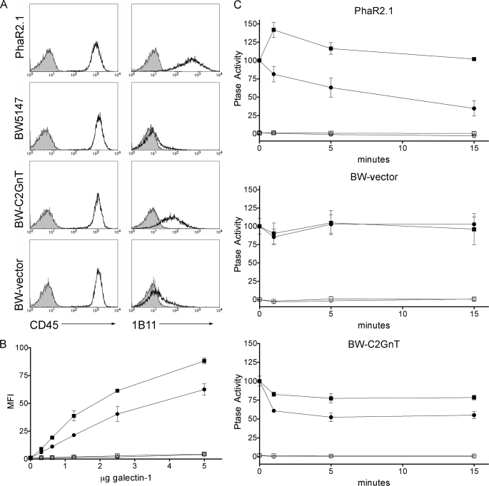
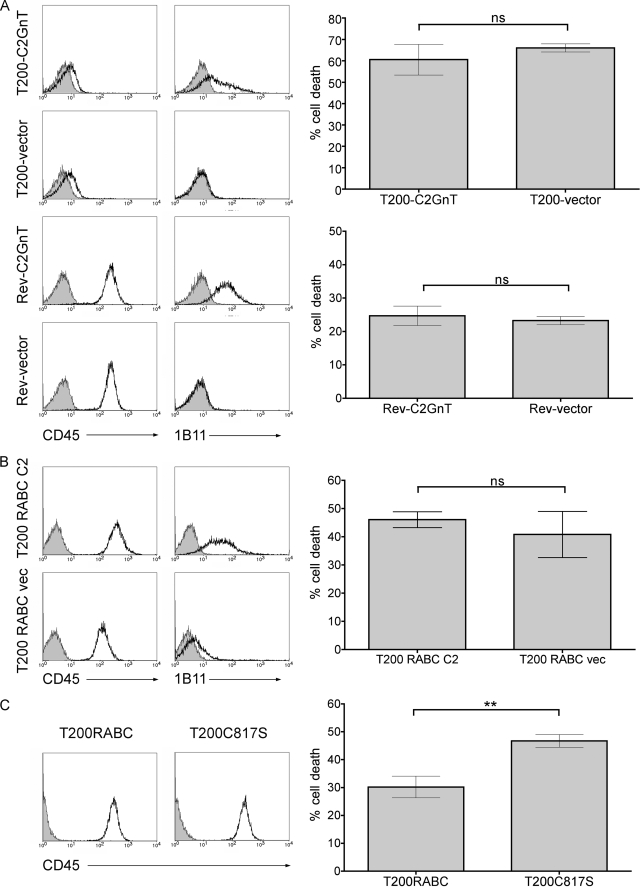
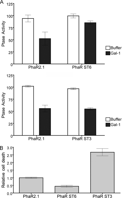
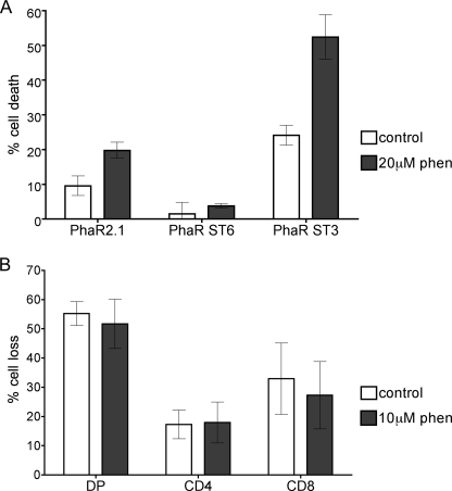
Galectin-1, a beta-galactoside-binding protein highly expressed in the thymus, induces apoptosis of specific thymocyte subsets and activated T cells. Galectin-1 binds to N- and O-glycans on several glycoprotein receptors, including CD7, CD43, and CD45. Here we show that galectin-1 signaling through CD45, which carries both N- and O-glycans, is regulated by CD45 isoform expression, core 2 O-glycan formation and the balance of N-glycan sialylation. Regulation of galectin-1 T cell death by O-glycans is mediated through CD45 phosphatase activity. While galectin-1 signaling in cells expressing low molecular weight isoforms of CD45 requires expression of core 2 O-glycans (high affinity ligands for galectin-1), galectin-1 signaling in cells expressing a high molecular weight isoform of CD45 does not require core 2 O-glycans, suggesting that a larger amount of core 1 O-glycans (low affinity ligands for galectin-1) is sufficient to overcome lack of core 2 O-glycans. Furthermore, regulation of galectin-1 signaling by alpha2,6-sialylation of N-glycans is not solely dependent on CD45 phosphatase activity and can be modulated by the relative expression of enzymes that attach sialic acid in an alpha2,6- or alpha2,3-linkage. Thus, N- and O-glycans modulate galectin-1 T cell death by distinct mechanisms, and different glycosylation events can render thymocytes susceptible or resistant to galectin-1.


Figures

FIGURE 1.
FIGURE 1.
Differential glycosylation of murine thymocyte subsets. A, identification of thymocyte subsets. Thymocytes were stained with CD4-FITC and CD8-PE. Representative gating to identify DN (CD4- CD8-), DP (CD4+ CD8+), CD4 (CD4+ CD8-), and CD8 (CD4- CD8+) cells is shown. B, DP thymocytes bear asialo O-glycans. Thymocytes were stained with CD4-FITC, CD8-PE, and PNA-biotin. Lectin binding was detected with streptavidin-PerCP. Thymocytes were gated on DN, DP, CD4, and CD8 subpopulations and PerCP MFI of each subset determined. C, SP thymocytes bear abundant α2,6-linked sialic acid. Cells were stained and gated as above, except that SNA-biotin binding was detected. D, SP thymocytes bear abundant α2,3-linked sialic acid. Cells were stained and gated as above, except that MAA-biotin binding was detected. In B–D, data are mean MFI ± S.E. of cells from four animals.
FIGURE 2.
FIGURE 2.
Derivation of cell lines used in this study. The PhaR2.1 cell line is a derivative of BW5147 that has spontaneously re-expressed C2GnT. The T200 cell line is a derivative of BW5147 that lacks CD45. The Rev1.1 cell line is a derivative of the T200 cell line that has spontaneously re-expressed the extracellular and transmembrane regions of CD45.
FIGURE 3.
FIGURE 3.
Expression of core 2 O-glycans increases galectin-1 binding and is required for galectin-1 inhibition of CD45 tyrosine phosphatase activity in cells expressing CD45RB. A, expression of C2GnT adds core 2 O-glycans to T cell surface glycoproteins. PhaR2.1, BW5147, BW-C2GnT, or BW-vector cells were stained with CD45-FITC or 1B11-PE (open) or isotype control (filled). 1B11 binding to BW-C2GnT cells demonstrates core 2 O-glycan addition. B, BW-C2GnT (squares) or BW-vector (circles) cells were bound by biotinylated galectin-1 for 10 min with (open) or without (filled) 50 mm lactose. Bound galectin-1 was detected with streptavidin-FITC, and MFI was determined. Data are mean fluorescence, corrected for autofluorescence, ± S.D. of duplicate samples. C, 1 × 10⁶ PhaR2.1, BW-vector, or BW-C2GnT cells were incubated with 20 μm galectin-1 (circles) or buffer control (squares) for the shown times. Cells were then washed and lysed, and phosphatase activity was measured with (open) or without (filled) bpV(phen). Data are mean ± S.D. of triplicate samples.
FIGURE 4.
FIGURE 4.
CD45 regulation of galectin-1 susceptibility requires the intracellular phosphatase domain, and CD45 isoform usage determines the requirement for core 2 O-glycans. A, T cells lacking the intracellular domain of CD45 do not require C2GnT expression for galectin-1 susceptibility. Left, T200 or Rev1.1 cells were transfected with cDNA encoding C2GnT or vector alone, and the presence of core 2 O-glycans determined by staining with 1B11-PE (open) or isotype control (filled). CD45 expression was determined with CD45-FITC (open) or isotype control (filled). Histograms are of representative clones. Right, T200-C2GnT, T200-vector, Rev-C2GnT, or Rev-vector cells were treated with galectin-1 or buffer control, and cell death was analyzed by staining with annexin-V-FITC and 7-AAD. T200 and Rev1.1 cells were susceptible to galectin-1 death, and cells with or without core 2 O-glycans had comparable levels of cell death. Data are mean ± S.D. of at least duplicate samples, and representative of at least four experiments. B, expression of core 2 O-glycans does not enhance galectin-1 susceptibility in T cells expressing CD45RABC. Left, T200 cells were transfected with CD45RABC and C2GnT (T200 RABC C2) or CD45RABC and vector control (T200 RABC vec), and core 2 O-glycan expression was determined as in A. Histograms are of representative clones. Right, cell death assays were performed as in A. Data are mean ± S.D. of triplicate samples. C, loss of CD45 phosphatase activity enhances T cell susceptibility to galectin-1. Left, T200 cells transfected with CD45RABC (T200RABC) or the inactive phosphatase mutant CD45RABC-C817S (T200C817S) were stained for CD45 as above. Right, while cells expressing CD45RABC were sensitive to galectin-1, complete loss of CD45 phosphatase activity in the C817S mutant enhanced susceptibility to galectin-1. Cell death assays were performed as above. Data are mean ± S.E. of triplicate samples from three independent experiments. Significance was determined by Student's t test.
FIGURE 5.
FIGURE 5.
ST6Gal-I and ST3Gal-III expression modulates the balance of sialic acid linkages on T cells to regulate galectin-1 binding. A, ST6Gal-I (PhaR ST6) and ST3Gal-III (PhaR ST3) were expressed in PhaR2.1 cells and cell surface sialylation was determined by reactivity with SNA-biotin or MAA-biotin, detected by streptavidin-FITC. Cells expressing ST6Gal-I had increased SNA binding and reduced MAA binding, and cells expressing ST3Gal-III had increased MAA binding and reduced SNA binding, indicating that the enzymes compete for acceptor substrates. Data are mean fluorescence intensity ± 95% CI for representative samples of three independent assays. B, overexpression of ST6Gal-I increased α2,6-linked sialic acid on CD45, while expression of ST3Gal-III reduced α2,6-linked sialic acid on CD45. Lysates of PhaR2.1, PhaR ST6, and PhaR ST3 cells were immunoprecipitated with anti-CD45 (45) or control IgG (C). Precipitates were probed with SNA-biotin, stripped, and reprobed with anti-CD45 to confirm equal loading. C, galectin-1 binding to bi-antennary N-glycans is regulated by addition of α2,6-linked sialic acid. Galectin-1 binding to a glycan microarray was performed, and binding to bi-antennary complex N-glycans decorated with different terminal sialic acid linkages was determined. Relative binding to a bi-antennary complex N-glycan with two α2,3 (column A), one α2,3 and one α2,6 (columns B and C), or two α2,6 (column D)-linked sialic acids. Data are mean relative fluorescence units (RFU) ± S.E. of six replicate determinations. Statistical significance was measured by one-way analysis of variance with Bonferroni's multiple comparison post-test. D, alteration of sialic acid balance on the cell surface results in change in galectin-1 binding. PhaR2.1 (■, □), PhaR ST6 (▾, ▿), or PhaR ST3 (●, ○) were incubated with indicated concentration of biotinylated galectin with (open) or without (filled) 50 mm lactose for 10 min, washed, and stained with streptavidin-FITC. Data are mean fluorescence intensity (corrected for autofluorescence) ± S.D. of duplicate samples.
FIGURE 6.
FIGURE 6.
Overexpression of ST6Gal-I, but not ST3Gal-III, inhibits galectin-1-induced reduction of CD45 phosphatase and death. A, PhaR2.1, PhaR ST6, and PhaR ST3 cells were incubated with 20 μm galectin-1 or buffer control for 15 min prior to determination of tyrosine phosphatase activity of cell lysates. Data are mean ± S.D. of triplicate samples. B, PhaR2.1, PhaR ST6, or PhaR ST3 cells were incubated with 20 μm galectin-1 or buffer control. % cell death was normalized to buffer control, and then PhaR ST6 and PhaR ST3 death were normalized relative to PhaR2.1 cell death. Data are mean ± S.E. of triplicate samples from seven independent experiments.
FIGURE 7.
FIGURE 7.
Inhibition of tyrosine phosphatase activity cannot overcome the block to galectin-1 death in T cells expressing high levels of α2,6-linked sialic acid. A, tyrosine phosphatase inhibitor bpV(phen) increases galectin-1 death of PhaR2.1 and PhaR ST3 cells, but does not affect PhaR ST6 cells. Phar2.1, PhaR ST6, and PhaR ST3 cells were treated with 20 μm bpV(phen) or buffer control for 3 h, then incubated with 20 μm galectin-1 or buffer control for an additional 4.5 h. Data are mean ± S.D. of triplicate samples. B, treatment with bpV(phen) does not increase galectin-1-induced death in SP thymocytes. Total thymocytes were incubated with 10 μm bpV(phen) and followed by 20 μm galectin-1 as above. Cells were stained with 7-AAD, anti-CD4, and anti-CD8, and gated for DP, CD4, and CD8 populations. Data are mean ± S.D. of triplicate samples from two independent experiments.
FIGURE 8.
FIGURE 8.
Removal of sialic acid from thymocytes renders SP thymocytes sensitive to galectin-1. A, thymocytes were treated with 100 units/ml C. perfringens neuraminidase or buffer control for 1 h. Cells were stained with CD4-FITC and CD8-PE, and SNA-biotin and streptavidin PerCP, and gated for DN, DP, CD4, and CD8 subpopulations. SNA binding is reduced on CD4 and CD8 cells after neuraminidase treatment. Data are mean fluorescence intensity ± S.D. of cells from two animals. B, loss of α2,6-linked sialic acid enhances galectin-1 susceptibility of CD4 and CD8 SP thymocytes. Cells were treated with neuraminidase or buffer control as above, followed by 20 μm galectin-1 or buffer control for 4.5 h. Cells were stained with 7-AAD, anti-CD4, and anti-CD8, and gated for DN, DP, CD4, and CD8 subpopulations. Cell loss from each subpopulation was determined. Data are mean ± S.D. of triplicate samples.

Similar articles

Cited by

References

    1. Hogquist K. A., Baldwin T. A., Jameson S. C. (2005) Nat. Rev. Immunol. 5, 772–782 - PubMed
    1. Baum L. G., Pang M., Perillo N. L., Wu T., Delegeane A., Uittenbogaart C. H., Fukuda M., Seilhamer J. J. (1995) J. Exp. Med. 181, 877–887 - PMC - PubMed
    1. Liu S. D., Whiting C. C., Tomassian T., Pang M., Bissel S. J., Baum L. G., Mossine V. V., Poirier F., Huflejt M. E., Miceli M. C. (2008) Blood 112, 120–130 - PMC - PubMed
    1. Liu S. D., Tomassian T., Bruhn K. W., Miller J. F., Poirier F., Miceli M. C. (2009) J. Immunol. 182, 5283–5295 - PubMed
    1. Perillo N. L., Pace K. E., Seilhamer J. J., Baum L. G. (1995) Nature 378, 736–739 - PubMed

Publication types

MeSH terms