. 2009 Dec 8;106(49):20776-81.
doi: 10.1073/pnas.0906998106. Epub 2009 Nov 17.

The anti-apoptotic protein HAX-1 is a regulator of cardiac function

Affiliations

The anti-apoptotic protein HAX-1 is a regulator of cardiac function

Wen Zhao et al. Proc Natl Acad Sci U S A. .

Abstract

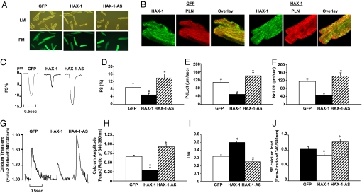
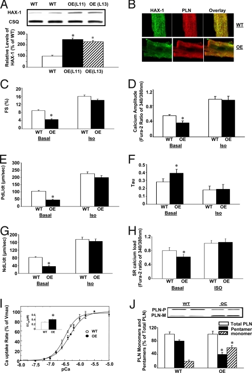
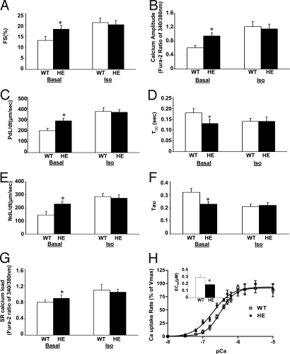
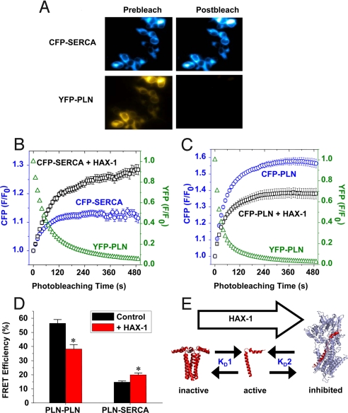
The HS-1 associated protein X-1 (HAX-1) is a ubiquitously expressed protein that protects cardiomyocytes from programmed cell death. Here we identify HAX-1 as a regulator of contractility and calcium cycling in the heart. HAX-1 overexpression reduced sarcoplasmic reticulum Ca-ATPase (SERCA2) pump activity in isolated cardiomyocytes and in vivo, leading to depressed myocyte calcium kinetics and mechanics. Conversely, downregulation of HAX-1 enhanced calcium cycling and contractility. The inhibitory effects of HAX-1 were abolished upon phosphorylation of phospholamban, which plays a fundamental role in controlling basal contractility and constitutes a key downstream effector of the beta-adrenergic signaling cascade. Mechanistically, HAX-1 promoted formation of phospholamban monomers, the active/inhibitory units of the calcium pump. Indeed, ablation of PLN rescued HAX-1 inhibition of contractility in vivo. Thus, HAX-1 represents a regulatory mechanism in cardiac calcium cycling and its responses to sympathetic stimulation, implicating its importance in calcium homeostasis and cell survival.


Conflict of interest statement

The authors declare no conflict of interest.

Figures

Fig. 1.
Fig. 1.
Effects of acute HAX-1 overexpression or downregulation on rat cardiomyocyte contractile function. Adenoviruses with sense and anti-sense mouse HAX-1 gene insertion were generated according to standard procedures. An adenovirus expressing GFP was used as a control. Rat adult cardiomyocytes were isolated and infected with Ad.GFP (GFP), Ad.HAX-1 (HAX-1), and Ad.HAX-1-antisense (HAX-1-AS). (A) Representative images of infected cardiomyocytes under light microscopy (LM, upper panel) and fluorescence microscopy (FM, lower panel). (B) Immunofluorescence analysis of HAX-1 localization in GFP and HAX-1 infected cardiomyocytes, using PLN as a SR marker. To investigate the contractile function, myocytes were infected with GFP, HAX-1, and HAX-1-AS for 24 h and contractility was subsequently recorded. (C) Representative curves of myocyte contraction under basal conditions. (D) Percentage of fractional shortening (FS%), (E) rate of contraction (PdL/dt, μm/s), and (F) rate of relaxation (NdL/dt, μm/s) under basal conditions. For the calcium transients, cells were loaded with fura-2 for 30 min, and then calcium transients were recorded and analyzed. (G) Representative curves of calcium transients under basal conditions. (H) Calcium amplitude, indicated as the fura-2 ratio of 340/380 nm; (I) Tau under basal conditions. For SR calcium load indicating by caffeine-induced calcium release from SR, 10 mM caffeine was added to the buffer. (J) Peak of SR calcium load, indicated by the fura-2 ratio of 340/380 nm. n = 4–5 hearts (6–8 cells/heart) for each group. Data are mean ± SEM. *, P < 0.05, compared to Ad.GFP group.
Fig. 2.
Fig. 2.
Generation and characterization of cardiac-specific HAX-1 overexpressing mice. HAX-1 transgenic mice were generated, using mouse HAX-1 cDNA under the control of an α-MHC promoter and human growth hormone polyA (HGH polyA). (A) Quantitative immunoblotting of heart homogenates from HAX-1 overexpressors (OE) and wild-type (WT). (B) Immunofluorescence analysis of HAX-1 localization in cardiomyocytes from WT and OE mice, using PLN as a SR marker. To analyze the contractile function, cardiomyocytes from OE or age-matched WT mice were isolated; contractility and calcium transients were recorded and analyzed under basal conditions (Basal) and upon isoproterenol stimulation (Iso, 100 nM). (C, E, and G) Percentage of fractional shortening (FS%), rate of contraction (PdL/dt, μm/s) and rate of relaxation (NdL/dt, μm/s). (D) Calcium amplitude, indicated by fura-2 ratio of 340/380 nm. (F) Tau. (H) Peak of SR calcium load, indicated by the fura-2 ratio of 340/380 nm. (I) Initial rates of oxalate-supported SR Ca uptake in cardiac homogenates from WT (○) and OE (●) mice. (Inset) The EC50 of SERCA for calcium in OE and WT hearts. Values were normalized to Vmax values. (J) Representative immunoblots (upper panel) and relative protein levels (lower panel) of PLN monomers and pentamers in WT and OE heart homogenates. Data are mean ± SEM. n = 6–8 hearts for each group (for C–H, 8–10 cells/heart). *, P < 0.05, compared to wild-type hearts.
Fig. 3.
Fig. 3.
Effect of HAX-1 deficiency on cardiomyocyte contractile function. Cardiomyocytes from HAX-1 heterozygous mice (HE) or age-matched wild-type (WT) were isolated; contractility and calcium transients, as well as caffeine induced calcium release from SR were recorded and analyzed. (A, C. and E) Percentage of fractional shortening (FS%), rate of contraction (PdL/dt, μm/s) and rate of relaxation (NdL/dt, μm/s). (B) Calcium amplitude, indicated by the fura-2 ratio of 340/380 nm. (D) Time to 50% decay of calcium (T50, s). (F) Tau. (G) Peak of SR calcium load, indicated by the fura-2 ratio of 340/380 nm. A–G are all under basal conditions (Basal) and upon isoproterenol stimulation (Iso). (H) Initial rates of oxalate-supported SR calcium uptake in hearts from WT (□) and HE (■) mice. (H, inset) The EC50 of SERCA for calcium in HE and WT hearts. Data are mean ± SEM. n = 4–6 hearts for each group (for A–G, 8–10 cells/heart). *, P < 0.05, compared to wild-type hearts.
Fig. 4.
Fig. 4.
Effect of HAX-1 on PLN-PLN and PLN-SERCA fluorescence resonance energy transfer (FRET). (A) Fluorescence microscopy showed that acceptor-selective photobleaching of YFP-PLN fluorescence resulted in an increase in CFP-SERCA fluorescence, indicating FRET. (B) Image quantification showed CFP-SERCA fluorescence (blue circles) increased exponentially as YFP-PLN (green triangles) was photobleached. The magnitude of CFP-SERCA fluorescence enhancement was greater in cells cotransfected with HAX-1 (black squares), suggesting increased FRET compared to control. (C) CFP-PLN fluorescence (blue circles) increased as YFP-PLN (green triangles) was bleached, indicating intrapentameric FRET. The magnitude of the CFP-PLN fluorescence enhancement was smaller in cells cotransfected with HAX-1 (black squares), suggesting reduced FRET. (D) Summary of FRET microscopy results. Coexpression of HAX-1 caused a 32% decrease in overall FRET between CFP-PLN and YFP-PLN, and a 35% increase in overall FRET between CFP-SERCA and YFP-PLN. Data are mean ± SEM, *, P < 0.01, compared with control. (E) A model for HAX-1 regulation of cardiac calcium handling. HAX-1 shifts the PLN pentamer/monomer equilibrium toward the active, monomeric species, and promotes formation of the PLN-SERCA regulatory complex.
Fig. 5.
Fig. 5.
Effect of HAX-1 overexpression on cardiomyocyte contractile performance in the absence of PLN. Cardiomyocytes were isolated from HAX-1 overexpression in the PLN knockout background (OE/KO), PLN knockout (PLN KO), and HAX-1 overexpression (HAX-1 OE) mice as well as age-matched wild-type (WT). Contractility and calcium transients as well as caffeine-induced SR calcium release from SR were recorded and analyzed. (A) Percentage of fractional shortening (FS%); (C) Rate of contraction (PdL/dt, μm/s); and (E) Rate of relaxation (NdL/dt, μm/s). (B) Calcium amplitude, indicated by fura-2 ratio of 340/380 nm. (D) Peak of SR calcium load, indicated by the fura-2 ratio of 340/380 nm. A–E are all under basal conditions. Data are mean ± SEM, n = 4–5 hearts (8–10 cells/heart) for each group. (F) Initial rates of oxalate-supported SR calcium uptake, and (G) EC50 of calcium uptake in heart homogenates from WT, HAX-1 OE, PLN KO and OE/KO mice. Data are mean ± SEM. n = 6–8 hearts for each group. *, P < 0.05, compared to WTs. ^, P < 0.05, compared to HAX-1 OEs.

Similar articles

Cited by

References

    1. Dash R, et al. Interactions between phospholamban and beta-adrenergic drive may lead to cardiomyopathy and early mortality. Circulation. 2001;103:889–896. - PubMed
    1. Haghighi K, et al. Superinhibition of sarcoplasmic reticulum function by phospholamban induces cardiac contractile failure. J Biol Chem. 2001;276:24145–24152. - PubMed
    1. Zhai J, et al. Cardiac-specific overexpression of a superinhibitory pentameric phospholamban mutant enhances inhibition of cardiac function in vivo. J Biol Chem. 2000;275:10538–10544. - PubMed
    1. MacLennan DH, Kranias EG. Phospholamban: A crucial regulator of cardiac contractility. Nat Rev Mol Cell Biol. 2003;4:566–577. - PubMed
    1. Vafiadaki E, et al. Phospholamban interacts with HAX-1, a mitochondrial protein with anti-apoptotic function. J Mol Biol. 2007;367:65–79. - PubMed

Publication types

MeSH terms