. 2009 Dec 1;106(48):20394-8.
doi: 10.1073/pnas.0909954106. Epub 2009 Nov 17.

Differential requirements of MHC and of DCs for endogenous proliferation of different T-cell subsets in vivo

Affiliations

Differential requirements of MHC and of DCs for endogenous proliferation of different T-cell subsets in vivo

Jeong-su Do et al. Proc Natl Acad Sci U S A. .

Abstract

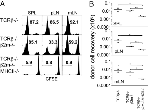
T cells transferred into severe lymphopenic hosts undergo rapid proliferation known as "endogenous proliferation" that are distinct from conventional homeostatic proliferation. Unlike homeostatic proliferation, cytokines, such as IL-7 are dispensable, yet TCR:MHC interaction is essential for this process to occur. However, cell types inducing the proliferation have not formally been addressed. In this study, we report that CD11c+ conventional DCs play irreplaceable roles in inducing endogenous proliferation of both naive and memory phenotype CD4 T cells via TCR-MHC II interaction. By contrast, CD8 T-cell endogenous proliferation was independent of MHC I or CD11c+ DCs. Interestingly, MHC II was necessary to support naive CD8 T-cell proliferation within MHC I-deficient hosts. Depletion of both B cells and DCs was sufficient to abrogate the proliferation of naive but not of memory CD8 T cells. These results suggest that depending on the T-cell lineages, as well as the differentiation status, different mechanisms control endogenous proliferation, revealing in vivo complexity of T-cell proliferation under lymphopenic conditions.


Conflict of interest statement

The authors declare no conflict of interest.

Figures

Fig. 1.
Fig. 1.
Roles of MHC in naive T-cell EP. (A) CFSE labeled naive Thy1.1 T cells were transferred into groups of TCRβ−/−, MHC II−/− TCRβ−/−, and β2m−/− TCRβ−/− recipients. CFSE profiles were examined 7 days after the transfer. The results shown are representative of six individually tested recipients. (B and C) Lethally irradiated TCRβ−/− mice were reconstituted with BM cells from TCRβ−/−, MHC II−/− TCRβ−/−, and β2m−/− TCRβ−/− donor mice. CFSE labeled naive Thy1.1 T cells were then transferred into the reconstituted recipients after 6 weeks of BM transfer. CFSE profiles were examined 7 days post transfer. Total donor cell recovery from the spleen was examined 16 days post transfer. The mean ± SD of donor T-cell proportion that fully diluted the CFSE (EP) is indicated. (D) MHC I−/− CD8 T cells were generated by bone marrow reconstitution. Naive CD44low MHC I−/− CD8 T cells isolated from the reconstituted recipients were CFSE labeled and transferred into groups of β2m−/− TCRβ−/− recipients. Shown are the CFSE profiles of TCRβ+/CD8+ gated cells determined 7 days post transfer. The mean ± SD of donor T-cell proportion that fully diluted the CFSE (EP) is indicated.
Fig. 2.
Fig. 2.
CD11c+ DC dependent CD4 T-cell EP. Groups of TCRβ−/− and DTR Tg TCRβ−/− mice were transferred with Thy1.1 naive T cells and treated with DTX and/or PDCA Ab as described in Methods. CFSE profiles of Thy1.1 donor T cells from the indicated tissues were examined 5 days after the transfer. The results shown are representative of four to six individually tested mice from two independent experiments. The mean ± SD of donor T-cell proportion that fully diluted the CFSE (EP) is indicated.
Fig. 3.
Fig. 3.
Role of B cells during naive T-cell EP following DC depletion. (A) Groups of DTR Tg TCRβ−/− mice were injected with 2B8 or 18B12 Abs. Six days later the recipients received CFSE labeled Thy1.1 naive T cells and then were subsequently treated with DTX or PBS (days −1 and 2). Recipients were killed 5 days after T-cell transfer. CFSE profiles in the indicated tissues were determined. The mean ± SD of donor T-cell proportion that fully diluted the CFSE (EP) is indicated. (B) Groups of DTR Tg TCRβ−/− mice were injected with 2B8, 18B12, or DTX, as described above. At day 0, 1 × 10⁶ Thy1.1 naive CD8 T cells were transferred into the treated recipients. At day 5 after the transfer surface activation markers (Ly6C, CD44, CD122, and CD127) of Thy1.1+ donor CD8 T cells were determined by FACS analysis. The histograms shown are representative of 3–10 individually tested recipients. The mean ± SD of Ly6Chigh (%), CD44high (MFI), CD122 (MFI), and CD127 (MFI) are indicated. (C and D) Cells from the indicated tissues of the groups described in (B) were stimulated with PMA/Ionomycin and cytokine expression was determined by intracellular staining. Shown are the mean ± SD of three to 10 individually tested mice. *, P < 0.05; **, P < 0.01; ***, P < 0.001.
Fig. 4.
Fig. 4.
MHC II-dependent CD8 T-cell EP. CFSE labeled naive Thy1.1 CD8 T cells were transferred (1 × 10⁶ per recipient) into groups of TCRβ−/−, TCRβ−/− β2m−/−, and TCRβ−/− β2m−/− MHC II−/− recipients. The recipients were killed 7 days post transfer and CFSE profiles of the donor CD8 T cells were determined by FACS analysis. (A) Histogram profiles shown are representative of two to three individually tested mice. (B) Total Thy1.1 CD8 T cells were enumerated. *, P < 0.05; **, P < 0.01; ***, P < 0.001
Fig. 5.
Fig. 5.
Role of APCs during memory T-cell EP following DC depletion. Groups of DTR Tg TCRβ−/− mice were injected with 2B8 or 18B12 Abs. Six days later the recipients received CFSE labeled CD44high CD8 T cells and then were subsequently treated with DTX or PBS (days −1 and 2). Recipients were killed 5 days after T-cell transfer and CFSE profiles were examined. Shown are CFSE profile representatives of three individually tested mice. The mean ± SD of donor T-cell proportion that fully diluted the CFSE (EP) is indicated.

References

    1. Kieper WC, et al. Recent immune status determines the source of antigens that drive homeostatic T cell expansion. J Immunol. 2005;174:3158–3163. - PubMed
    1. Min B, Yamane H, Hu-Li J, Paul WE. Spontaneous and homeostatic proliferation of CD4 T cells are regulated by different mechanisms. J Immunol. 2005;174:6039–6044. - PubMed
    1. Tan JT, et al. IL-7 is critical for homeostatic proliferation and survival of naive T cells. Proc Natl Acad Sci USA. 2001;98:8732–8737. - PMC - PubMed
    1. Guimond M, et al. Interleukin 7 signaling in dendritic cells regulates the homeostatic proliferation and niche size of CD4+ T cells. Nat Immunol. 2009;10:149–157. - PMC - PubMed
    1. Tan JT, et al. Interleukin (IL)-15 and IL-7 jointly regulate homeostatic proliferation of memory phenotype CD8+ cells but are not required for memory phenotype CD4+ cells. J Exp Med. 2002;195:1523–1532. - PMC - PubMed

Publication types

LinkOut - more resources