. 2009 Dec 15;69(24):9228-35.
doi: 10.1158/0008-5472.CAN-09-1950.

Extracellular signal-regulated kinase signaling pathway regulates breast cancer cell migration by maintaining slug expression

Affiliations

Extracellular signal-regulated kinase signaling pathway regulates breast cancer cell migration by maintaining slug expression

Haoming Chen et al. Cancer Res. .

Abstract

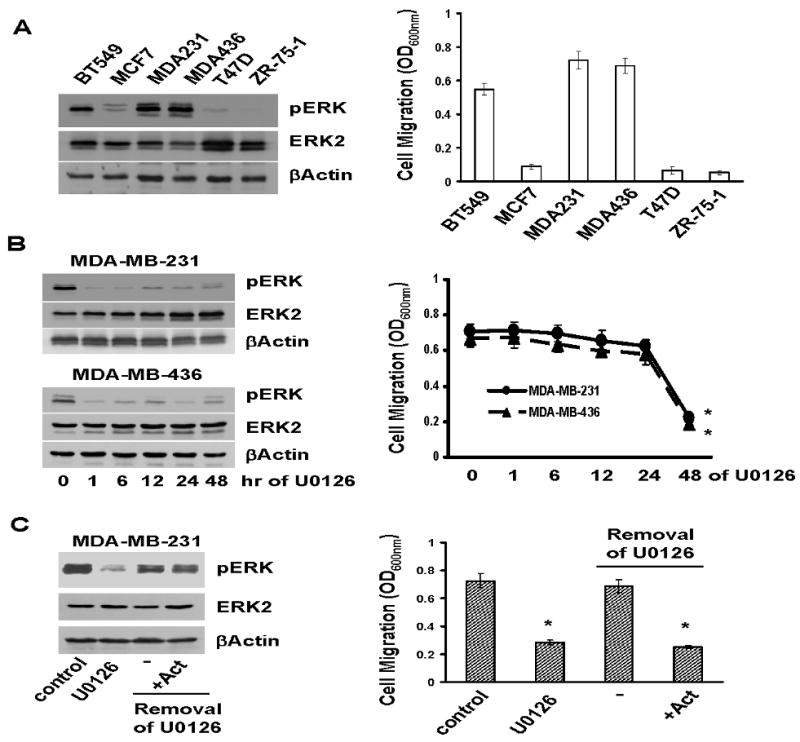
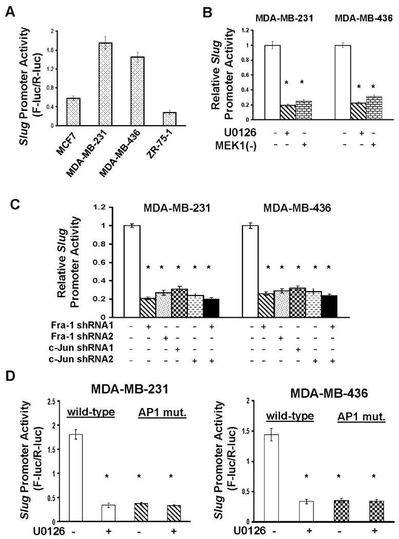
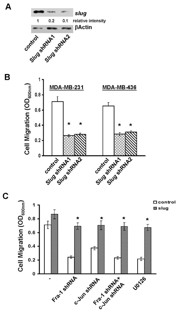
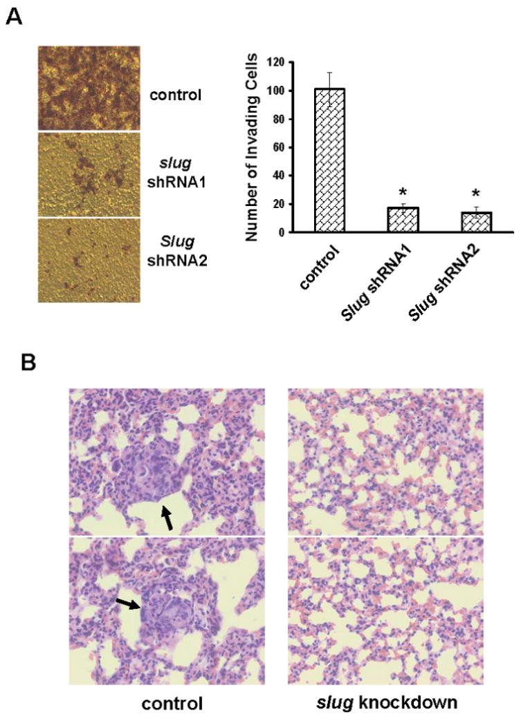
Cell migration is a critical step in cancer cell invasion. Recent studies have implicated the importance of the extracellular signal-regulated kinase (ERK) signaling pathway in cancer cell migration. However, the mechanism associated with ERK-regulated cell migration is poorly understood. Using a panel of breast cancer cell lines, we detected an excellent correlation between ERK activity and cell migration. Interestingly, we noticed that a 48-hour treatment with U0126 [specific mitogen-activated protein/ERK kinase (MEK)-1/2 inhibitor] was needed to significantly inhibit breast cancer cell migration, whereas this inhibitor blocked ERK activity within 1 hour. This observation suggests that ERK-dependent gene expression, rather than direct ERK signaling, is essential for cell migration. With further study, we found that ERK activity promoted the expression of the activator protein-1 (AP1) components Fra-1 and c-Jun, both of which were necessary for cell migration. Combination of U0126 treatment and Fra-1/c-Jun knockdown did not yield further reduction in cell migration than either alone, indicating that ERKs and Fra-1/c-Jun act by the same mechanism to facilitate cell migration. In an attempt to investigate the role of Fra-1/c-Jun in cell migration, we found that the ERK-Fra-1/c-Jun axis regulated slug expression in an AP1-dependent manner. Moreover, the occurrence of U0126-induced migratory inhibition coincided with slug reduction, and silencing slug expression abrogated breast cancer cell migration. These results suggest an association between ERK-regulated cell migration and slug expression. Indeed, cell migration was not significantly inhibited by U0126 treatment or Fra-1/c-Jun silencing in cells expressing slug transgene. Our study suggests that the ERK pathway regulates breast cancer cell migration by maintaining slug expression.


Figures

Fig.1
Fig.1. ERK-dependent gene expression is required for breast cancer cell migration
A. A portion of overnight-cultured BT549, MCF7, MDA-MB-231, MDA-MB-436, T47D and ZR-75-1 cells were lysed and lysates subjected to immunoblotting to detect phosphor-ERK, ERK2 and βactin with the respective antibodies. The remaining portion of cells was analyzed for cell migration using Transwells as described in “Materials and Methods”. Data are means ± S.E. B. MDA-MB-231 and MDA-MB-436 cells were treated with 5μM U0126 for various times. A portion of cells was analyzed for the levels of phosphor-ERK, ERK2 and βactin and the remaining portion analyzed for cell migration. Data are means ± S.E (*, p < 0.005 vs 0-hr). C. MDA-MB-231 cells were treated with 5μM U0126 for 2 days, then washed with serum-free medium three times to remove U0126, and incubated in the absence or the presence of 2 μg/ml actinomycin for 4 h. A portion of cells were analyzed for phosphor-ERK, ERK2 and βactin levels and the remaining portion assayed for cell migration. Data are means ± S.E. (*, p < 0.005 vs control).
Fig.2
Fig.2. ERK-Fra-1/c-Jun axis is involved in cell migration
A. BT549, MCF7, MDA-MB-231, MDA-MB-436, T47D and ZR-75-1 cells were lysed and lysates subjected to immunoblotting to detect Fra-1, c-Jun, Fra-2 and βactin with the respective antibodies. B. MDA-MB-231 and MDA-MB-436 cells were treated with 5μM U0126 for various times and then lysed for immunoblotting to detect Fra-1, c-Jun, Fra-2 and βactin. C. MDA-MB-231 and MDA-MB-436 cells were either infected with control lentiviral vector (luciferase shRNA) or vector containing Fra-1, c-Jun shRNA or both. Some Fra-1/c-Jun shRNA expressing cells were further treated with 5μM U0126 for 2 days. Cells were detached with 10 mM EDTA and then assayed for cell migration as described in “Materials and Methods”. Data are means ± S.E. (*, p < 0.005 vs control; #, p < 0.01 vs control).
Fig.3
Fig.3. ERK-Fra-1/c-Jun axis regulates slug expression
A. MDA-MB-231 and MDA-MB-436 cells were treated with 5μM U0126 for various times and then lysed for immunoblotting to detect slug and βactin with the respective antibodies. B. MDA-MB-231 cells were infected with lentiviral vector containing luciferase shRNA (control), Fra-1 or c-Jun shRNA for 4 days and then lysed for immunoblotting to detect slug and βactin.
Fig.4
Fig.4. Slug is expressed in an AP1-dependent manner
A. MCF7, MDA-MB-231, MDA-MB-436 and ZR-75-1 cells were transfected with the slug promoter luciferase reporter plasmid for 2 days, then lysed and cell lysates measured for luciferase activity as described in “Materials and Methods”. Data are means ± S.E. B. MDA-MB-231 and MDA-MB-436 cells were transfected with the slug promoter plasmid for 1 day and 5μM U0126 then added to cells for another 2 days. In a parallel expreriment, the slug promoter plasmid was co-transfected into cells with dominant negative MEK1 expression vector [MEK1(-)] for 3 days. Cells were lysed and cell lysates analyzed for luciferase activity. Data are means ± S.E. (*, p < 0.005 vs control). C. The slug promoter plasmid was transfected into MDA-MB-231 and MDA-MB-436 cells that expressed Fra-1 or c-Jun shRNA or both for 2 days. Cells were lysed and cell lysates analyzed for luciferase activity. Data are means ± S.E. (*, p < 0.005 vs control). D. MDA-MB-231 and MDA-MB-436 cells were transfected with the slug promoter plasmid or plasmid containing slug promoter with mutation in AP1 consensus site for 1 day and 5μM U0126 added to cells for another 2 days. Cells were lysed and cell lysates measured for luciferase activity. Data are means ± S.E. (*, p < 0.005 vs cells with wild-type promoter/no U0126).
Fig.5
Fig.5. Slug is essential for ERK-Fra-1/c-Jun axis-regulated cell migration
A. MDA-MB-231 cells were infected with lentiviral vector containing luciferase shRNA (control) or slug shRNA for 4 days and then lysed for immunoblotting to detect slug and βactin with the respective antibodies. B. MDA-MB-231 and MDA-MB-436 cells were infected with control (luciferase shRNA) or slug shRNA-containing lentiviral vector for 4 days, then detached with 10 mM EDTA and assayed for cell migration as described in “Materials and Methods”. Data are means ± S.E. (*, p < 0.005 vs control). C. MDA-MB-231 cells were infected with empty lentiviral vector (control) or vector encoding murine slug cDNA for 4 days. Control cells or cells with stable murine slug cDNA expression were further infected with lentiviral vector containing luciferase shRNA, Fra-1 shRNA or c-Jun shRNA or both for 4 days. In a parallel experiment, control and murine slug cDNA-expressing cells were treated with 5μM U0126 for 2 days. Cells were detached and analyzed for cell migration. Data are means ± S.E. (*, p < 0.005 vs control).
Fig.6
Fig.6. Slug is essential for in vitro invasion and lung metastasis
A. Control or slug-knockdown MDA-MB-231 cells were added into invasion chambers and allowed to invade for 24 hrs. The cells on the undersurface of chambers were stained and visualized under microscope. Data are means ± S.E. (*, p < 0.001 vs control). B. The lungs from mice receiving either control or slug-knockdown MDA-MB-231 cells for 8 weeks were fixed and sectioned. The sections were subjected to H&E staining and visualized under microscope. The arrows point at metastatic lesions and the images are in 40× magnification.

References

    1. Friedl P, Wolf K. Tumour-cell invasion and migration: diversity and escape mechanisms. Nature Rev Cancer. 2003;3:362–74. - PubMed
    1. Wehrle-Haller B, Imhof BA. Actin, microtubules and focal adhesion dynamics during cell migration. Int J Biochem Cell Biol. 2003;35:39–50. - PubMed
    1. Javelaud D, Laboureau J, Gabison E, Verrecchia F, Mauviel A. Disruption of basal JNK activity differentially affects key fibroblast functions important for wound healing. J Biol Chem. 2003;278:24624–8. - PubMed
    1. Meadows KN, Bryant P, Vincent PA, Pumiglia KM. Activated Ras induces a proangiogenic phenotype in primary endothelial cells. Oncogene. 2004;23:192–200. - PubMed
    1. Yamauchi J, Chan JR, Shooter EM. Neurotrophin 3 activation of TrkC induces Schwann cell migration through the c-Jun N-terminal kinase pathway. Proc Natl Acad Sci. 2003;100:14421–6. - PMC - PubMed

Publication types

MeSH terms