. 2009 Dec 1;69(23):8932-40.
doi: 10.1158/0008-5472.CAN-08-3873. Epub 2009 Nov 17.

Human bone marrow-derived mesenchymal stem cells for intravascular delivery of oncolytic adenovirus Delta24-RGD to human gliomas

Affiliations

Human bone marrow-derived mesenchymal stem cells for intravascular delivery of oncolytic adenovirus Delta24-RGD to human gliomas

Raymund L Yong et al. Cancer Res. .

Abstract

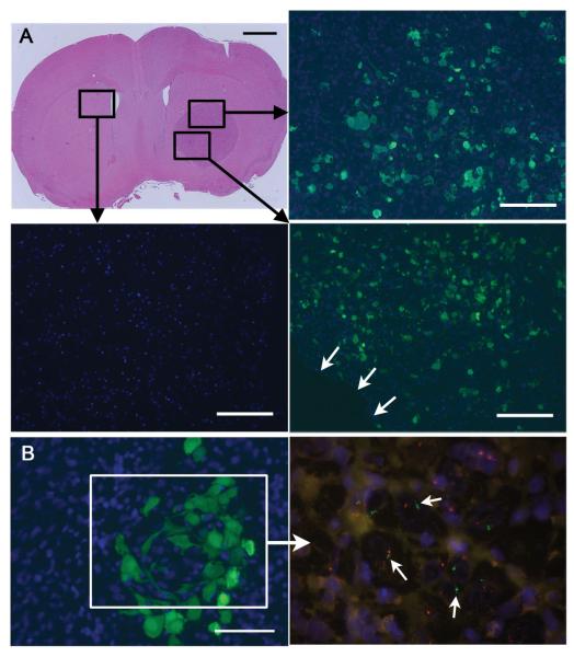
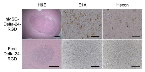
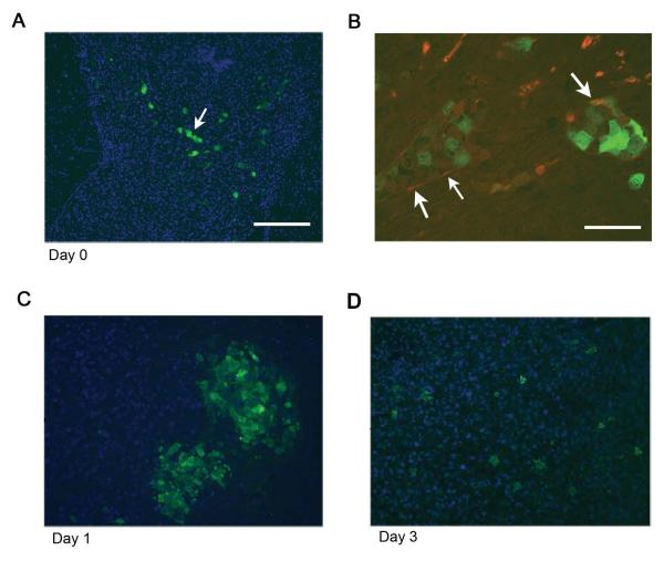
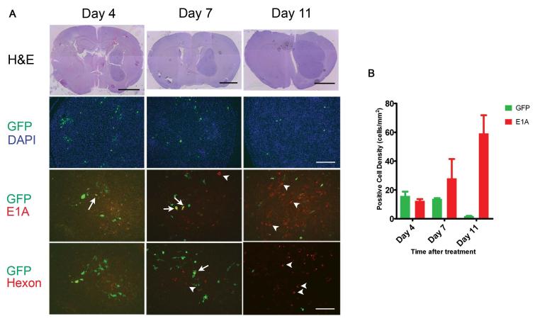
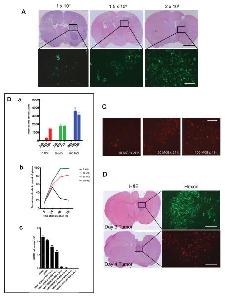
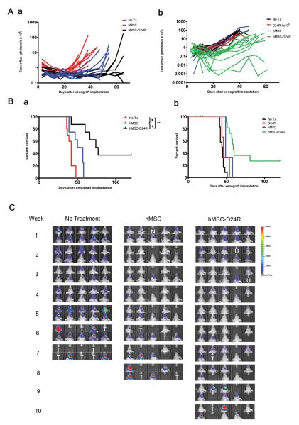
Delta24-RGD is an infectivity-augmented, conditionally replicative oncolytic adenovirus with significant antiglioma effects. Although intratumoral delivery of Delta24-RGD may be effective, intravascular delivery would improve successful application in humans. Due to their tumor tropic properties, we hypothesized that human mesenchymal stem cells (hMSC) could be harnessed as intravascular delivery vehicles of Delta24-RGD to human gliomas. To assess cellular events, green fluorescent protein-labeled hMSCs carrying Delta24-RGD (hMSC-Delta24) were injected into the carotid artery of mice harboring orthotopic U87MG or U251-V121 xenografts and brain sections were analyzed by immunofluorescence for green fluorescent protein and viral proteins (E1A and hexon) at increasing times. hMSC-Delta24 selectively localized to glioma xenografts and released Delta24-RGD, which subsequently infected glioma cells. To determine efficacy, mice were implanted with luciferase- labeled glioma xenografts, treated with hMSC-Delta24 or controls, and imaged weekly by bioluminescence imaging. Analysis of tumor size by bioluminescence imaging showed inhibition of glioma growth and eradication of tumors in hMSC-Delta24-treated animals compared with controls (P < 0.0001). There was an increase in median survival from 42 days in controls to 75.5 days in hMSC-Delta24-treated animals (P < 0.0001) and an increase in survival beyond 80 days from 0% to 37.5%, respectively. We conclude that intra-arterially delivered hMSC-Delta24 selectively localize to human gliomas and are capable of delivering and releasing Delta24-RGD into the tumor, resulting in improved survival and tumor eradication in subsets of mice.


Figures

Figure 1
Figure 1
A, Representative mouse implanted with U87MG in the right frontal lobe. One million GFP-labeled hMSCs were injected into the right carotid artery on day 7. The brain was extracted for histology on day 11 (scale bar 1 mm). The boxed areas were subjected to immunofluorescence using anti-GFP antibodies to reveal the distribution of hMSCs (green cells) within the xenograft. Blue = DAPI nuclear dye. B, A cluster of GFP-labeled hMSCs of genotype XY is seen within a U87MG xenograft of genotype XO after intracarotid delivery (scale bar 50 μm). The boxed area shows FISH on an adjacent section to reveal Y chromosomes (green, arrows) within the area where hMSCs are present. Outside this area, only cells containing X chromosomes (red) are seen.
Figure 2
Figure 2
hMSC-Delta24 produces tumor infections while free Delta-24-RGD does not. Mice were treated as described in the text and brain sections immunostained with anti-E1A or anti-hexon antibodies. Viral E1A protein and hexon protein were seen throughout hMSCs-Delta24-treated brains (brown cytoplasmic stain) but not in controls treated with free virus. Additionally, E1A and hexon were absent in brains treated with uninfected hMSCs and phosphate-buffered saline (not shown). Scale bars 500 μm H&E sections, and 50 μm immunostained sections.
Figure 3
Figure 3
A, Brain section from a mouse sacrificed immediately after hMSC delivery demonstrating the presence of GFP-labeled hMSCs organized in linear patterns (arrows), suggesting arrival via tumor blood vessels. Scale bar 200 μm. B, Section from a separate mouse stained with antibodies against endothelial marker CD31 and GFP. GFP-positive cells (green) are hMSCs and CD31 positive cells (red) are endothelial cells. Arrows show area where red endothelial cells surround green hMSCs. Scale bar 50 μm. C, D, Mice were implanted with U87MG and then treated with hMSC-Delta24-RGD-AdGFP. Analysis of brains on days 1 and 3 by fluorescence microscopy after staining anti-GFP antibody (green) revealed hMSCs arranged in clusters on day 1 (C). By day 3, hMSCs had dispersed within the xenografts (D). Blue=DAPI.
Figure 4
Figure 4
A, Immunofluorescence microscopy was used to track hMSCs and Delta-24-RGD over time after intracarotid delivery to U87MG xenografts. Brains were harvested 4, 7, and 11 days after treatment. Sections were cut and stained with H&E (Upper row). Sections were double immunostained with FITC-labeled anti-GFP antibody (green) and with Texas Red-labeled anti-E1A antibody (third row, red) or Texas Red-labeled anti-hexon antibody (lowest row, red). Sections were also stained with DAPI (second row). Green cells indicate hMSCs that are not expressing viral proteins; yellow cells indicate hMSCs that are expressing viral proteins; red cells indicate U87 tumor cells expressing viral proteins. Arrows indicate examples of hMSC double-expressing GFP and E1A (yellow cells). Arrow-heads indicate examples of E1A-expressing U87MG cells (red cells). The pattern indicates progression from green to yellow to red cells, suggesting movement of virus from hMSCs to glioma cells. H&E scale bars 2mm, GFP/DAPI scale bar 200μm, and EIA and hexon scale bars 100μm. B, Graph showing the density of GFP-positive cells (i.e., hMSCs) and adenoviral protein-expressing cells (i.e. cells supporting viral replication) within U87MG xenografts over the course of the experiment depicted in a. (p=0.0006 for interaction, 2-way ANOVA).
Figure 5
Figure 5
Optimization of delivery of Delta-24-RGD to U87MG xenografts via hMSCs. A, An increased number of delivered hMSCs correlates with an increased number of engrafted hMSCs. Mice were implanted with 5 × 10⁵ U87MG cells in the right frontal lobe and allowed to grow for 7 days. 1.0, 1.5, or 2 × 10⁶ GFP-labled-hMSCs were injected into the carotid artery. Mice were sacrificed 4 days later. H&E scale bar 2 mm, hMSC-GFP scale bar 100 μm. B, (a) hMSCs were infected with Delta-24-RGD at 10, 50, or 100 MOI and allowed to incubate for 24, 48, or 72 hours. Cells were collected along with media and the titer of Delta-24-RGD was measured to determine the number of infectious units released. The highest number of infectious units was obtained using 100 MOI incubated for 48 hours. Experiment performed in triplicate. (b) hMSCs were plated in serum-free media and allowed to synchronize. At 0 hours, cells were infected with Delta-24-RGD at 0, 10, 50, or 100 MOI. At the indicated time points, cell cycle analysis was performed. Results are the percentage of cells in S-phase or pseudo-S-phase. All hMSCs infected with 50 or 100 MOI exhibited evidence of viral DNA replication by 48 hours. (c) hMSCs were infected with Delta-24-RGD at the indicated MOIs, then washed and plated in the upper wells of a Transwell plate (1×10⁴cells/well). U87MG cells were plated in the bottom wells (3×10⁴cells/well) and incubate for 7 days, after which U87MG cells were counted. Killing was seen with MOIs ≥ 10 and incubation times of ≥24hrs. C, Mice were implanted with U87MG cells and treated with hMSCs-Delta24 at the indicated MOIs and incubation times. Four days later, brains were analyzed by immunofluorescence for hexon protein expression (red cells). The 50MOI × 24h and 100MOI × 48h incubations produced equivalent infections. Scale bar 100μm. D, Mice were implanted with U87MG cells and injected IC with 1.5×10⁶ hMSCs-Delta24 (50 MOI×24h incubation) 3 (upper) or 4 (lower) days later. On day 7 after delivery of hMSCs, xenografts were analyzed for hexon protein expression (green in top panel and red in bottom panel). H&E scale bar 1 mm, hexon scale bar 200 μm.
Figure 6
Figure 6
A, Photon flux of xenografts over 60 days for two independent experiments in which nude mice were implanted with luminescent U87MG xenografts (U87MG-GL in a and U87MG-LucNeo in b, 5 × 10⁵ cells). In each experiment, 10 mice were left untreated and 10-20 mice underwent two treatments with “empty” hMSCs or hMSCs-Delta24 (1.5×10⁶ cells, 50MOI×24h incubation). Mice were imaged weekly. Total photonic flux was measured from fixed regions of interest encompassing the entire head. Each line represents efflux from a single animal. Slower growth is seen with hMSC-Delta24 treatment. B, Survival data corresponding to the two experiments presented in A. In Experiment 1 (a) median survival was 77 days in treated animals compared to 42 days in untreated controls and 53.5 days in animals given “empty” hMSCs. Among treated animals, 37.5% lived beyond 80 days. * P < 0.01 and ** P = 0.0002, log-rank test. In Experiment 2 (b), median survival was 60 days in treated animals compared to 45 days in untreated controls. 36% of animals lived beyond 80 days. *** P < 0.0001, log-rank test. C, Representative bioluminescence images over the time course of the experiment depicted in a. Individual mice were tracked. N, no animal. U, death unrelated to tumor. T, tumor-related death.

Similar articles

Cited by

References

    1. Stupp R, Mason WP, van den Bent MJ, et al. Radiotherapy plus concomitant and adjuvant temozolomide for glioblastoma. N Engl J Med. 2005;352:987–96. - PubMed
    1. Fueyo J, Gomez-Manzano C, Alemany R, et al. A mutant oncolytic adenovirus targeting the Rb pathway produces anti-glioma effect in vivo. Oncogene. 2000;19:2–12. - PubMed
    1. Fueyo J, Alemany R, Gomez-Manzano C, et al. Preclinical characterization of the antiglioma activity of a tropism-enhanced adenovirus targeted to the retinoblastoma pathway. J Natl Cancer Inst. 2003;95:652–60. - PubMed
    1. Krasnykh V, Dmitriev I, Mikheeva G, Miller CR, Belousova N, Curiel DT. Characterization of an adenovirus vector containing a heterologous peptide epitope in the HI loop of the fiber knob. J Virol. 1998;72:1844–52. - PMC - PubMed
    1. Dmitriev I, Krasnykh V, Miller CR, et al. An adenovirus vector with genetically modified fibers demonstrates expanded tropism via utilization of a coxsackievirus and adenovirus receptor-independent cell entry mechanism. J Virol. 1998;72:9706–13. - PMC - PubMed

Publication types

MeSH terms