

Inhibition of the p38 kinase suppresses the proliferation of human ER-negative breast cancer cells
- PMID: 19920204
- PMCID: PMC2830975
- DOI: 10.1158/0008-5472.CAN-09-1636
Inhibition of the p38 kinase suppresses the proliferation of human ER-negative breast cancer cells
Abstract
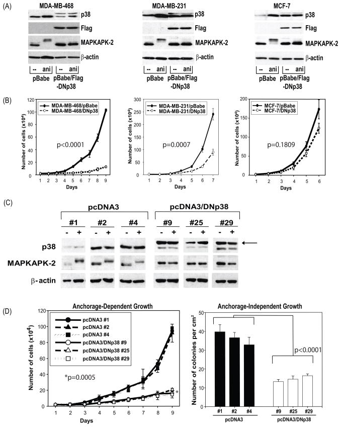
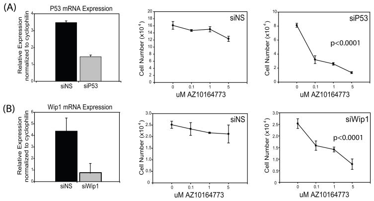
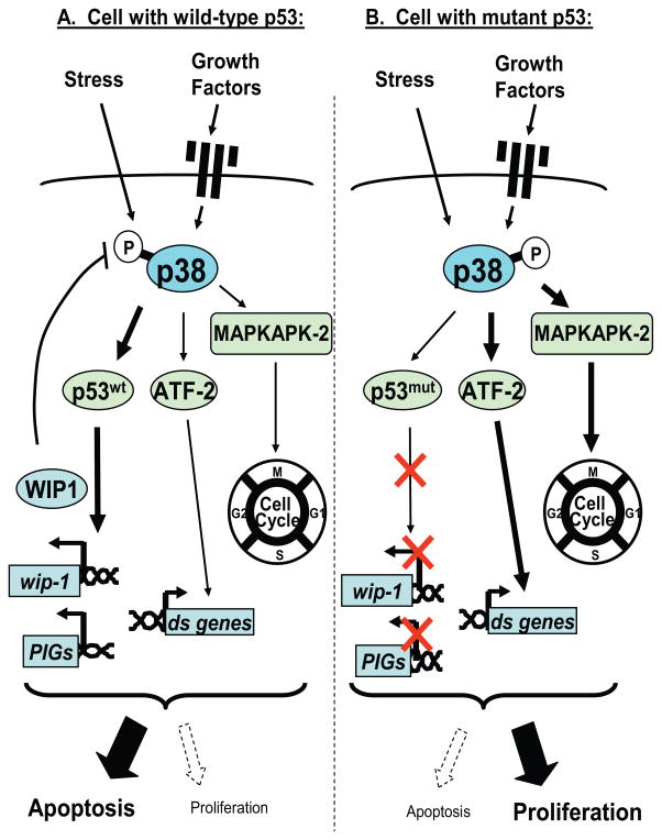
p38 kinases are members of the mitogen-activated protein kinase family that transduce signals from various environmental stresses, growth factors, and steroid hormones. p38 is highly expressed in aggressive and invasive breast cancers. Increased levels of activated p38 are markers of poor prognosis. In this study, we tested the hypothesis that blockade of p38 signaling would inhibit breast cancer cell proliferation. We studied breast cancer cell proliferation and cell cycle regulation upon p38 blockade by using three independent approaches: dominant-negative (DN) constructs, small interfering RNA (siRNA), and small molecule inhibitors. p38alpha and p38delta are the most abundant isoforms expressed by all examined human breast tumors and breast cancer cell lines. Expression of a DN p38 inhibited both anchorage-dependent and -independent proliferation of MDA-MB-468 cells. Silencing of p38alpha, but not p38delta, using siRNA suppressed MDA-MB-468 cell proliferation. Pharmacologic inhibitors of p38 significantly inhibited the proliferation of p53 mutant and ER-negative breast cancer cells. Whereas p38 has previously been considered as a mediator of stress-induced apoptosis, we propose that p38 may have dual activities regulating survival and proliferation depending on the expression of p53. Our data suggest that p38 mediates the proliferation signal in breast cancer cells expressing mutant but not wild-type p53. Because most ER-negative breast tumors express mutant p53, our results provide the foundation for future development of p38 inhibitors to target p38 for the treatment of p53 mutant and ER-negative breast cancers.
Figures


Similar articles
-
Overexpression of the wip1 gene abrogates the p38 MAPK/p53/Wip1 pathway and silences p16 expression in human breast cancers.Breast Cancer Res Treat. 2007 Mar;101(3):269-78. doi: 10.1007/s10549-006-9304-y. Epub 2006 Aug 9. Breast Cancer Res Treat. 2007. PMID: 16897432
-
Accumulation of tissue factor in endothelial cells induces cell apoptosis, mediated through p38 and p53 activation.Thromb Haemost. 2015 Aug;114(2):364-78. doi: 10.1160/TH14-09-0795. Epub 2015 Apr 23. Thromb Haemost. 2015. PMID: 25903973
-
Proteasome inhibitors induce a p38 mitogen-activated protein kinase (MAPK)-dependent anti-apoptotic program involving MAPK phosphatase-1 and Akt in models of breast cancer.Breast Cancer Res Treat. 2006 Nov;100(1):33-47. doi: 10.1007/s10549-006-9232-x. Epub 2006 Jun 29. Breast Cancer Res Treat. 2006. PMID: 16807678
-
Arsenic trioxide augments Chk2/p53-mediated apoptosis by inhibiting oncogenic Wip1 phosphatase.J Biol Chem. 2008 Jul 4;283(27):18969-79. doi: 10.1074/jbc.M800560200. Epub 2008 May 15. J Biol Chem. 2008. PMID: 18482988
-
Evaluating the role of p38 MAP kinase in growth of Werner syndrome fibroblasts.Ann N Y Acad Sci. 2010 Jun;1197:45-8. doi: 10.1111/j.1749-6632.2010.05195.x. Ann N Y Acad Sci. 2010. PMID: 20536832 Review.
Cited by
-
Anticancer effect of Tamarix gallica extracts on human colon cancer cells involves Erk1/2 and p38 action on G2/M cell cycle arrest.Cytotechnology. 2013 Dec;65(6):927-36. doi: 10.1007/s10616-013-9564-4. Epub 2013 Jun 26. Cytotechnology. 2013. PMID: 23801270 Free PMC article.
-
Sublethal concentrations of the platinum(II) complex [Pt(O,O'-acac)(gamma-acac)(DMS)] alter the motility and induce anoikis in MCF-7 cells.Br J Pharmacol. 2010 Jul;160(6):1362-77. doi: 10.1111/j.1476-5381.2010.00782.x. Br J Pharmacol. 2010. PMID: 20590627 Free PMC article.
-
Bioorthogonal Engineering of Bacterial Effectors for Spatial-Temporal Modulation of Cell Signaling.ACS Cent Sci. 2019 Jan 23;5(1):145-152. doi: 10.1021/acscentsci.8b00751. Epub 2018 Dec 27. ACS Cent Sci. 2019. PMID: 30693333 Free PMC article.
-
Inhibition of p38 MAPK sensitizes tumour cells to cisplatin-induced apoptosis mediated by reactive oxygen species and JNK.EMBO Mol Med. 2013 Nov;5(11):1759-74. doi: 10.1002/emmm.201302732. Epub 2013 Sep 24. EMBO Mol Med. 2013. PMID: 24115572 Free PMC article.
-
p38γ promotes breast cancer cell motility and metastasis through regulation of RhoC GTPase, cytoskeletal architecture, and a novel leading edge behavior.Cancer Res. 2011 Oct 15;71(20):6338-49. doi: 10.1158/0008-5472.CAN-11-1291. Epub 2011 Aug 23. Cancer Res. 2011. PMID: 21862636 Free PMC article.
References
-
- Society AC. Breast Cancer Facts and Figures 2009. Atlanta: American Cancer Society; 2009.
-
- Fisher B, Costantino JP, Wickerham DL, et al. Tamoxifen for prevention of breast cancer: report of the National Surgical Adjuvant Breast and Bowel Project P-1 Study. J Natl Cancer Inst. 1998;90:1371–88. - PubMed
-
- Cummings SR, Eckert S, Krueger KA, et al. The effect of raloxifene on risk of breast cancer in postmenopausal women: results from the MORE randomized trial. Multiple Outcomes of Raloxifene Evaluation. Jama. 1999;281:2189–97. - PubMed
-
- American CS. Estimated new cancer cases and deaths by gender. Atlanta: American Cancer Society; 2002.
-
- Tan AR, Swain SM. Adjuvant chemotherapy for breast cancer: an update. Semin Oncol. 2001;28:359–76. - PubMed
Publication types
MeSH terms
Substances
Grants and funding
LinkOut - more resources
Full Text Sources
Other Literature Sources
Medical
Research Materials
Miscellaneous
