. 2009 Nov;21(11):3535-53.
doi: 10.1105/tpc.109.070672. Epub 2009 Nov 17.

Definition of early transcriptional circuitry involved in light-induced reversal of PIF-imposed repression of photomorphogenesis in young Arabidopsis seedlings

Affiliations

Definition of early transcriptional circuitry involved in light-induced reversal of PIF-imposed repression of photomorphogenesis in young Arabidopsis seedlings

Pablo Leivar et al. Plant Cell. 2009 Nov.

Abstract

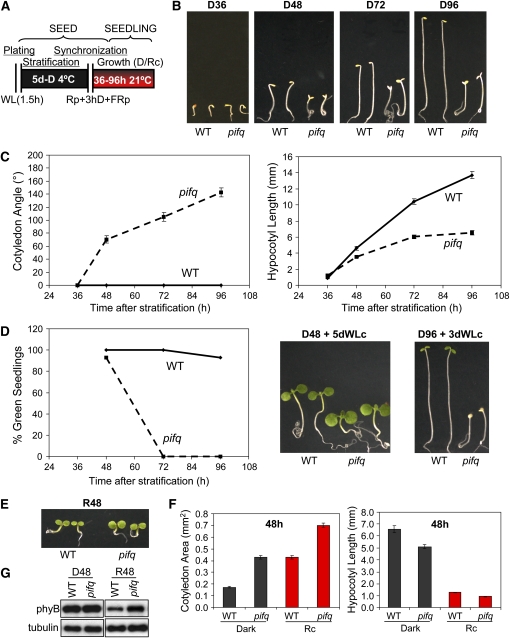
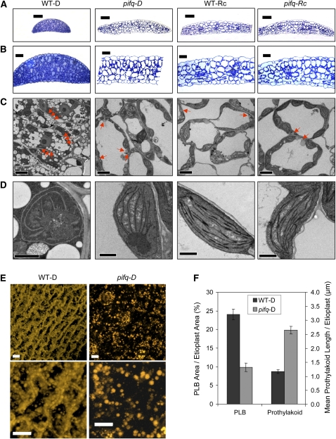
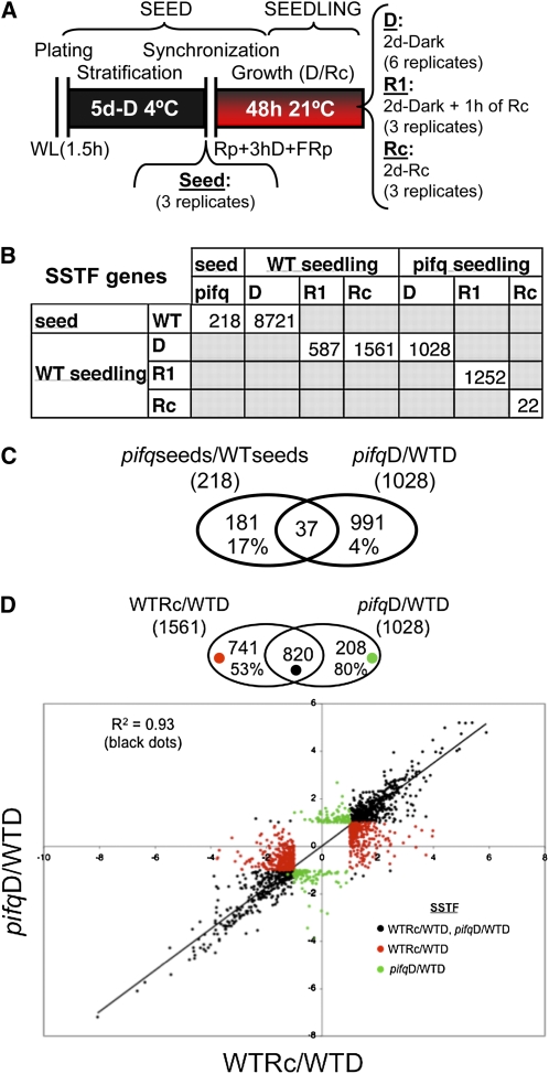
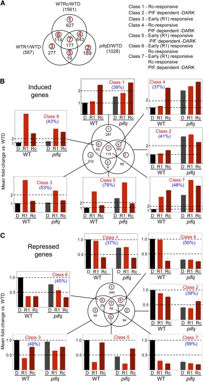
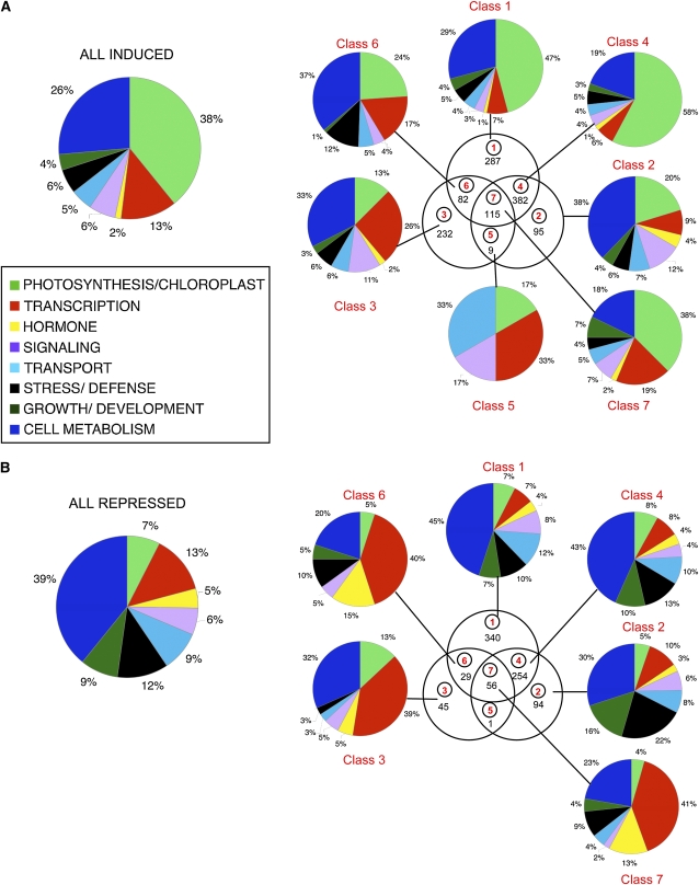
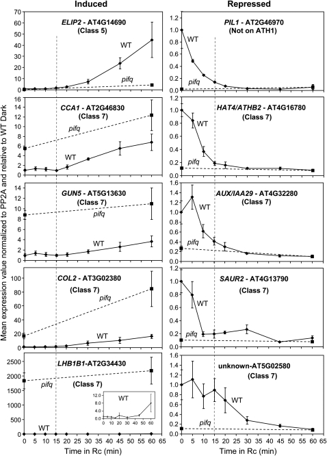
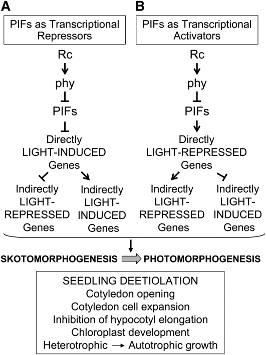
Light signals perceived by the phytochromes induce the transition from skotomorphogenic to photomorphogenic development (deetiolation) in dark-germinated seedlings. Evidence that a quadruple mutant (pifq) lacking four phytochrome-interacting bHLH transcription factors (PIF1, 3, 4, and 5) is constitutively photomorphogenic in darkness establishes that these factors sustain the skotomorphogenic state. Moreover, photoactivated phytochromes bind to and induce rapid degradation of the PIFs, indicating that the photoreceptor reverses their constitutive activity upon light exposure, initiating photomorphogenesis. Here, to define the modes of transcriptional regulation and cellular development imposed by the PIFs, we performed expression profile and cytological analyses of pifq mutant and wild-type seedlings. Dark-grown mutant seedlings display cellular development that extensively phenocopies wild-type seedlings grown in light. Similarly, 80% of the gene expression changes elicited by the absence of the PIFs in dark-grown pifq seedlings are normally induced by prolonged light in wild-type seedlings. By comparing rapidly light-responsive genes in wild-type seedlings with those responding in darkness in the pifq mutant, we identified a subset, enriched in transcription factor-encoding genes, that are potential primary targets of PIF transcriptional regulation. Collectively, these data suggest that the transcriptional response elicited by light-induced PIF proteolysis is a major component of the mechanism by which the phytochromes pleiotropically regulate deetiolation and that at least some of the rapidly light-responsive genes may comprise a transcriptional network directly regulated by the PIF proteins.


Figures

Figure 1.
Figure 1.
Time-Course Analysis of pifq Mutant Morphological Phenotypes in Dark and Light. (A) Schematic representation of the protocol used for seedling growth under true dark (Leivar et al., 2008b) and Rc conditions. Seeds were exposed to 1.5 h of white light (WL) during sterilization and plating and were placed for 5 d at 4°C in darkness (D) (stratification). A poststratification treatment consisting of a 5-min R pulse (Rp) followed by 3 h D incubation and a terminal 5-min far-red (FR) pulse (Rp+3 hD+FRp) was provided before incubation at 21°C in D or Rc (6.7 μmol/m2/s) for the indicated period. (B) Time-course analysis of the visible morphological phenotypes of pifq mutants grown in darkness for 36 to 96 h (D36-D96). Photos of representative wild-type and pifq mutant seedlings are shown. (C) Time-course analysis of cotyledon separation (left panel) and hypocotyl length (right panel) phenotypes of wild-type and pifq mutant seedlings grown in darkness. (D) Time-course analysis of the photobleaching phenotype of wild-type and pifq mutant seedlings grown in darkness for the indicated time before transferring them to continuous WL (WLc) for 3 to 5 d. The percentage of green seedlings was scored from at least 45 seedlings (left panel), and photos of representative wild-type and pifq mutant seedlings are shown (right panel). (E) Visible morphological phenotypes of pifq mutants grown in Rc for 48 h (R48). Photos of representative wild-type and pifq mutant seedlings are shown. (F) Quantification of the cotyledon area (left panel) and the hypocotyl length (right panel) of wild-type and pifq mutant seedlings grown in the dark or Rc for 48 h. (G) Immunoblot analysis of phyB protein levels in wild-type and pifq mutant seedlings grown in the dark or Rc for 48 h. Tubulin was used as loading control. A representative blot is shown. Quantification of replicated experiments is shown in Supplemental Figure 3 online. Data in (C) and (F) represent the mean and se of at least 20 seedlings.
Figure 2.
Figure 2.
Cellular and Subcellular Phenotypes of Dark-Grown pifq Seedlings Phenocopy Rc-Grown Wild Type. Wild-type and pifq mutant seedlings were grown in D or Rc for 48 h as in Figure 1A. (A) Toluidine blue staining of semithin (0.5 μm) cross sections of cotyledons. Micrographs were taken by light microscopy. Bars = 50 μm. (B) Higher-magnification micrographs of samples prepared as in (A). Bars = 20 μm. (C) Micrographs obtained by transmission electron microscopy of cross sections of cotyledons. High-pressure freezing and microwave processing methods were used for sample preparation, and several micrographs were taken for both methods. Representative images of the cell morphology of dark- and Rc-grown wild-type and pifq seedlings are shown. Red arrows indicate oil bodies. Bars = 5 μm. (D) Higher-magnification micrographs of samples prepared as in (C). Representative etioplasts (D grown) and chloroplasts (Rc grown) are shown for wild-type and pifq seedlings. Bars = 1 μm. (E) Nile red staining of wild-type and pifq mutant cotyledons grown in darkness. The cotyledons were stained with Nile Red (Greenspan et al., 1985; Siloto et al., 2006) and examined by confocal microscopy. Maximum intensity projections from 36 slices are shown. The bottom images are higher magnifications of the top ones. Bars = 10 μm. (F) Quantification of PLB area as a percentage of host etioplast area and mean prothylakoid length per etioplast. Data represent the mean and se of at least 76 etioplasts from several different transmission electron micrographs of wild-type and pifq mutant cotyledons grown in the dark. PLB area and prothylakoid length was measured using Image J.
Figure 3.
Figure 3.
Genetic Removal of PIFs Robustly Phenocopies in Darkness the Sustained Transcriptome Changes Elicited in Wild-Type Seedlings in Rc. (A) Schematic representation of the protocol for wild-type and pifq seed and seedling growth used for microarray-based transcriptome analysis. Samples were harvested directly at the end of the stratification period (seeds) or after 2 d of growth at 21°C (seedlings) under true dark conditions (D) or in Rc as described in Figure 1A. In addition, 2-d-old true dark–grown seedlings were irradiated with 7.5 μmol/m2/s of R for 1 h (R1). The number of biological replicates for each sample is indicated. (B) Number of differentially expressed genes that are defined as SSTF different in the pairwise comparisons indicated in the matrix. Expression data and primary analysis for the SSTF genes are reported in Supplemental Data Set 1 online. (C) Genes that are PIF regulated in both seed and dark-grown seedlings. Venn diagram shows pairwise comparison between SSTF genes differentially expressed in seeds (wild-type seed versus pifq-seed) and seedlings (WT-D versus pifq-D). The percentage of shared genes that are differentially expressed in each set is indicated. The list of shared genes (37) is provided in Supplemental Data Set 2 online. (D) Comparison of long-term Rc-responsive genes (WT-D versus WT-Rc) and PIF-regulated genes in dark-grown seedlings (WT-D versus pifq-D). Top: Venn diagram compares the SSTF differentially expressed genes in the two genotype growth treatment combinations. The number and percentage of shared genes in the comparison are indicated. Bottom: Scatterplot of log2 fold change values provides a quantitative measure of the correlation in responsiveness for each gene between the two genotype growth treatment combinations. Black dots in the scatterplot represent genes that are shared between the two combinations in the Venn diagram (top), whereas red and green dots represent genes that are specifically present in one of the combinations but not in the other. A trend line and the correlation coefficient for the shared genes (black dots) are indicated.
Figure 4.
Figure 4.
Genome-Wide Patterns of Rapidly Rc Light–Responsive and PIF-Regulated Gene Expression Reveal Pathway Convergence on Potential Target Genes. Three-way comparison of SSTF genes responding to 1 h Rc (WT-D versus WT-R1), long-term (2d)-Rc (WT-D versus WT-Rc), and to the pifq mutations in darkness (WT-D versus pifq-D). Classification of the genes as induced (B) or repressed (C) is based on the direction of the response, relative to WT-D, elicited by each genotype treatment combination. A small group of 25 genes in Classes 4 to 7 were designated as ambiguous as the direction of the response relative to WT-D differed between WT-R1, WT-Rc, and/or pifqD. These genes are listed in Supplemental Data Set 5 online and were excluded from the analysis shown in (B) and (C). The mean fold change in expression relative to WT-D (set at unity) for all genes in each class is shown in the bar graphs in (B) and (C). Error bars represent the mean standard error for the genes averaged for each genotype treatment combination. Percentage of genes having a G-box in the 3-kb upstream regulatory sequence is indicated in parenthesis. (A) Venn diagram showing comparison among all genes in the three different sets of SSTF differentially regulated genes (left panel). This comparison between genes responding to WT-R1, WT-Rc, and pifq-D resulted in the definition of seven classes of responsive genes (right panel) corresponding to the sectors of the diagram (circled numbers in red). The number of genes in each sector/class is indicated in black. (B) Induced genes. The number of induced genes in each class is indicated by the black numbers in each sector of the Venn diagram. The gene lists are in Supplemental Data Set 3 online. (C) Repressed genes. The number of repressed genes in each class is indicated by the black numbers in each sector of the Venn diagram. The gene lists are in Supplemental Data Set 4 online.
Figure 5.
Figure 5.
Functional Categories of Class 1 to 7 Induced and Repressed Genes. Induced (A) and repressed (B) genes in each class (1 to 7) were assigned separately to a functional category (color coded). This assignment was based on Gene Ontology annotations for biological and/or molecular function available at TAIR (http://www.Arabidopsis.org). The percentage of the total annotated genes within each class/sector was calculated after excluding the genes with unknown biological or molecular function. The distribution of all the induced (A) and all the repressed (B) genes is also shown for comparison (left).
Figure 6.
Figure 6.
Differing Patterns of Rapid Light Responsiveness Suggest Alternate Modes of PIF Regulation of Early Response Genes. Wild-type (solid curve) and pifq mutant (dashed curve) seedlings were grown for 2 d in the dark as indicated in Figure 3A and then exposed to Rc (7.5 μmol/m2/s) for increasing periods from 0 (dark control) to 60 min. Expression of the indicated genes was determined by qPCR, and PP2A was used as a normalization control as described (Shin et al., 2007). Data are presented relative to the mean of WT-D set at unity and represent the mean and se of three independent biological replicates. Vertical dashed line marks the 15-min Rc time point.
Figure 7.
Figure 7.
Simplified Schematic Model Depicting the Formal Alternate Direct and Indirect Control Modes Potentially Exercised by the PIF Proteins to Transcriptionally Activate or Repress Genes Regulating Seedling Deetiolation in Response to phy-Mediated Light Signals. The PIFs are shown as acting constitutively in darkness as either transcriptional repressors (A) or activators (B) of direct target genes. Light-activated phy molecules trigger reversal of each potential activity through induced proteolytic degradation of the PIF factors, thereby derepressing light-induced direct target genes (A) and inactivating light-repressed direct target genes (B). Regulatory genes (such as transcription factor genes) within this direct target category then act either positively or negatively on downstream genes in the transcriptional network, thereby indirectly inducing or repressing their expression in response to the light signal.

Similar articles

Cited by

References

    1. Al-Sady, B., Kikis, E.A., Monte, E., and Quail, P.H. (2008). Mechanistic duality of transcription factor function in phytochrome signaling. Proc. Natl. Acad. Sci. USA 105 2232–2237. - PMC - PubMed
    1. Al-Sady, B., Ni, W., Kircher, S., Schafer, E., and Quail, P.H. (2006). Photoactivated phytochrome induces rapid PIF3 phosphorylation prior to proteasome-mediated degradation. Mol. Cell 23 439–446. - PubMed
    1. Bae, G., and Choi, G. (2008). Decoding of light signals by plant phytochromes and their interacting proteins. Annu. Rev. Plant Biol. 59 281–311. - PubMed
    1. Bajracharya, D., and Schopfer, P. (1979). Effect of light on the development of glyoxysomal functions in the cotyledons of mustard (Sinapis alba L.) seedlings. Planta 145 181–186. - PubMed
    1. Bauer, D., Viczian, A., Kircher, S., Nobis, T., Nitschke, R., Kunkel, T., Panigrahi, K.C., Adam, E., Fejes, E., Schafer, E., and Nagy, F. (2004). Constitutive photomorphogenesis 1 and multiple photoreceptors control degradation of phytochrome interacting factor 3, a transcription factor required for light signaling in Arabidopsis. Plant Cell 16 1433–1445. - PMC - PubMed

Publication types

MeSH terms

Associated data