. 2009 Dec;119(12):3652-65.
doi: 10.1172/JCI39401. Epub 2009 Nov 16.

Live imaging reveals a biphasic mode of dissemination of Borrelia burgdorferi within ticks

Affiliations

Live imaging reveals a biphasic mode of dissemination of Borrelia burgdorferi within ticks

Star M Dunham-Ems et al. J Clin Invest. 2009 Dec.

Abstract

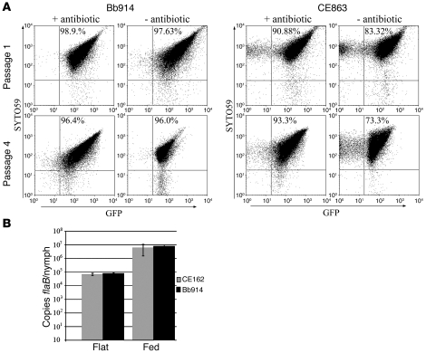
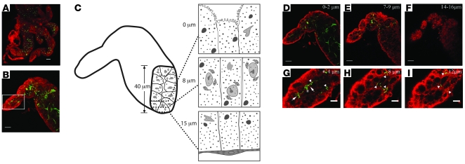
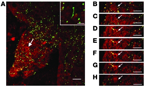
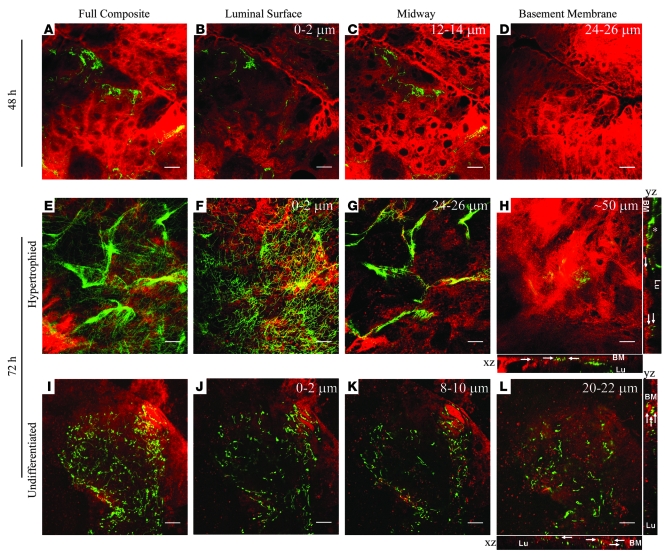
Lyme disease is caused by transmission of the spirochete Borrelia burgdorferi from ticks to humans. Although much is known about B. burgdorferi replication, the routes and mechanisms by which it disseminates within the tick remain unclear. To better understand this process, we imaged live, infectious B. burgdorferi expressing a stably integrated, constitutively expressed GFP reporter. Using isolated tick midguts and salivary glands, we observed B. burgdorferi progress through the feeding tick via what we believe to be a novel, biphasic mode of dissemination. In the first phase, replicating spirochetes, positioned at varying depths throughout the midgut at the onset of feeding, formed networks of nonmotile organisms that advanced toward the basolateral surface of the epithelium while adhering to differentiating, hypertrophying, and detaching epithelial cells. In the second phase of dissemination, the nonmotile spirochetes transitioned into motile organisms that penetrated the basement membrane and entered the hemocoel, then migrated to and entered the salivary glands. We designated the first phase of dissemination "adherence-mediated migration" and provided evidence that it involves the inhibition of spirochete motility by one or more diffusible factors elaborated by the feeding tick midgut. Our studies, which we believe are the first to relate the transmission dynamics of spirochetes to the complex morphological and developmental changes that the midgut and salivary glands undergo during engorgement, challenge the conventional viewpoint that dissemination of Lyme disease-causing spirochetes within ticks is exclusively motility driven.


Figures

Figure 1
Figure 1. Bb914 stably maintains a GFP reporter while retaining wild-type infectivity for ticks.
(A) Flow cytometric comparison of Bb914 (PflaB-gfp inserted into cp26) and CE863 (PflaB-gfp in the shuttle vector pCE323) (34) grown to mid-logarithmic phase following temperature shift in the presence or absence of antibiotics. The percentages of GFP+/SYTO59+ spirochetes are indicated in the upper-right-hand quadrant of each cytogram. Each flow cytogram is representative of 3 independent experiments. (B) Spirochete burdens of Bb914- and CE162-infected nymphs before and after feeding on naive C3H/HeJ mice. The infected nymphs used in these studies were generated by feeding naive larvae on syringe-inoculated C3H/HeJ mice as described in Methods. Data represent the means from 3 independent experiments; errors bars indicate SD.
Figure 2
Figure 2. B. burgdorferi are distributed throughout the midguts of flat nymphs.
(A) Low-power epifluorescence image of an isolated unfed midgut. (B) Composite confocal image showing the distribution of spirochetes through the full thickness (40 μm) of an unfed midgut. (C) Scheme used to acquire optical sections through an unfed Bb914-infected midgut and cartoon depiction of the cellular morphology at varying depths within the epithelium (modeled after ref. 46). (DF) 3-μm composite images of optical sections acquired (D) at the luminal surface, (E) midway through the epithelium, and (F) at the basement membrane. (GI) Digital enlargements of single, 1-μm optical sections of the boxed area in B acquired (G) at the luminal surface, (H) midway through the epithelium, and (I) at the most distal point where spirochetes were observed. Arrows, arrowheads, and asterisks indicate spirochete aggregates, intercellular spirochetes, and extracellular blebs, respectively; localization of spirochetes and blebs was determined by optical sectioning. Here and elsewhere, red staining denotes FM4-64; green staining denotes GFP. Scale bars: 50 μm (A); 25 μm (B, DF); 10 μm (GI).
Figure 3
Figure 3. Spirochetes shed myriad blebs during early feeding (24 hours after placement).
(A) Composite confocal image showing the distribution of spirochetes and numerous blebs through the full thickness of a midgut (45 μm); shown in the inset is a spirochete shedding blebs. Arrow indicates the cell in the consecutive confocal sections in BH. (BH) Localization of an intracellular bleb (arrow) present within A by consecutive 0.5-μm confocal sections acquired through the full depth (3.5 μm) of an epithelial cell. N, nucleus. Scale bars: 25 μm.
Figure 4
Figure 4. B. burgdorferi progress through the nymphal midgut during feeding in the form of epithelial cell–associated networks.
Representative confocal micrographs of midguts isolated from Bb914-infected nymphs at (AD) 48 and (EL) 72 hours after placement; EH and IL, respectively, show hypertrophied and undifferentiated regions of 72-hour-fed midguts. Composite images in A, E, and I depict the full thickness of the respective epithelial layers. 3-μm composite images show spirochetes (B, F, and J) at the luminal surface (C, G, and K) midway through the epithelium and (D, H, and L) at the basement membrane. Orthogonal views (y-z and x-z axes) for the 72-hour-fed midguts in H and L show that some spirochetes near the basement membrane (arrows) are in a head-on orientation. The positions of the lumen (Lu) and basement membrane (BM) in each orthogonal view are indicated. The asterisk in the y-z axis view of H denotes a spirochetal network. Scale bars: 25 μm.
Figure 5
Figure 5. Spirochetes advance around and between epithelial cells toward the basement membrane.
(AC) Representative silver-stained paraffin sections and (DF) FM4-64–labeled cryosections of midguts isolated from Bb914-infected nymphs at (A and D) 48 and (B, C, E, and F) 72 hours after placement on naive C3H/HeJ mice. (F) Digital enlargement of the boxed area in E. Representative examples of differentiated (dc) and undifferentiated cells (uc) are indicated. Arrows indicate spirochete aggregates on the epithelial surface (AC) or aggregates within the intercellular spaces (D). Asterisks in F indicate organisms in a head-on orientation near the basement membrane. Scale bars: 25 μm (AE); 5 μm (F).
Figure 6
Figure 6. Motility of B. burgdorferi in a gelatin matrix model system is inhibited by fed midgut.
Trajectory maps showing the motility of Borrelia in (A) BSK-II medium, (B) a gelatin matrix alone, (C) a gelatin matrix with a sample well containing fresh mouse blood, and (D) a gelatin matrix with a sample well containing a 72-hour-fed midgut from an uninfected nymph. The trajectory maps were generated from time-lapse epifluorescence images acquired adjacent to the sample well during the first minute of viewing using the MultiTrack plug-in of Image J (95).
Figure 7
Figure 7. Penetration of B. burgdorferi into the hemocoel is a rare event.
(A) A spirochete (arrow) traversing the basement membrane of an isolated midgut obtained 72 hours after placement on a naive C3H/HeJ mouse. The inset shows a digital enlargement of the penetrating spirochete. (B and C) Representative micrographs of (B) a spirochete and (C) a hemocyte containing GFP-positive material within hemolymph collected 72 hours after placement. Scale bars: 10 μm.
Figure 8
Figure 8. B. burgdorferi are present on salivary glands in low numbers and are observed only within types II and III acini.
Representative micrographs of salivary glands isolated from (A) flat or (BD) fed nymphs at 72 hours after placement on C3H/HeJ mice. Spirochetes (arrowheads) adhering to the surfaces of (B) type III acini and type I (inset). (C) Representative 10-μm composite confocal image showing spirochetes on the surfaces of and within types II and III acini. Spirochetes on the surface (arrowheads), below the surface near the basement membrane (filled arrows), between epithelial cells (open arrows), and within ducts (asterisks) are indicated. (D) Single view taken from a 3D reconstruction showing spirochetes on the surface of (arrowheads), penetrating into (asterisks), and within (arrows) a type III acinus. Note the size difference between type I acini (B, inset), which do not hypertrophy, and the hypertrophied types II and III acini in BD. Scale bars: 25 μm (AD); 5 μm (B, inset).

Similar articles

Cited by

References

    1. WHO. 2008. World health statistics. http://www.who.int/whosis/whostat/2008/en/index.html.
    1. Hill C.A., Kafatos F.C., Stansfield S.K., Collins F.H. Arthropod-borne diseases: vector control in the genomics era. Nat. Rev. Microbiol. 2005;3:262–268. doi: 10.1038/nrmicro1101. - DOI - PubMed
    1. Hemingway J., Beaty B.J., Rowland M., Scott T.W., Sharp B.L. The Innovative Vector Control Consortium: improved control of mosquito-borne diseases. Trends Parasitol. 2006;22:308–312. doi: 10.1016/j.pt.2006.05.003. - DOI - PubMed
    1. Sacks D., Kamhawi S. Molecular aspects of parasite-vector and vector-host interactions in leishmaniasis. Annu. Rev. Microbiol. 2001;55:453–483. doi: 10.1146/annurev.micro.55.1.453. - DOI - PubMed
    1. Barillas-Mury C., Kumar S. Plasmodium-mosquito interactions: a tale of dangerous liaisons. . Cell Microbiol. 2005;7:1539–1545. doi: 10.1111/j.1462-5822.2005.00615.x. - DOI - PubMed

Publication types

MeSH terms

Substances