. 2009 Dec;119(12):3573-85.
doi: 10.1172/JCI40202. Epub 2009 Nov 16.

Th22 cells represent a distinct human T cell subset involved in epidermal immunity and remodeling

Affiliations

Th22 cells represent a distinct human T cell subset involved in epidermal immunity and remodeling

Stefanie Eyerich et al. J Clin Invest. 2009 Dec.

Abstract

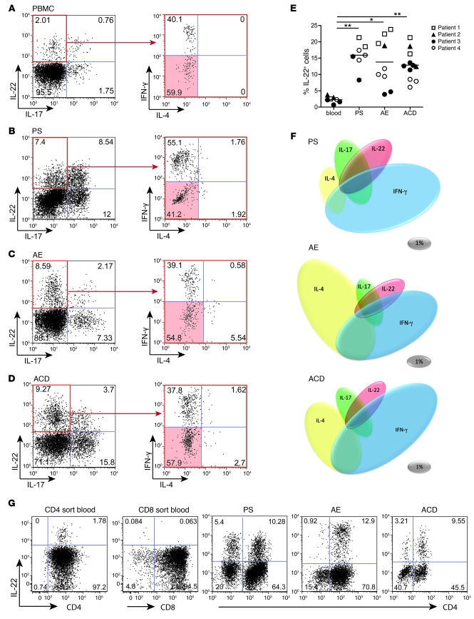
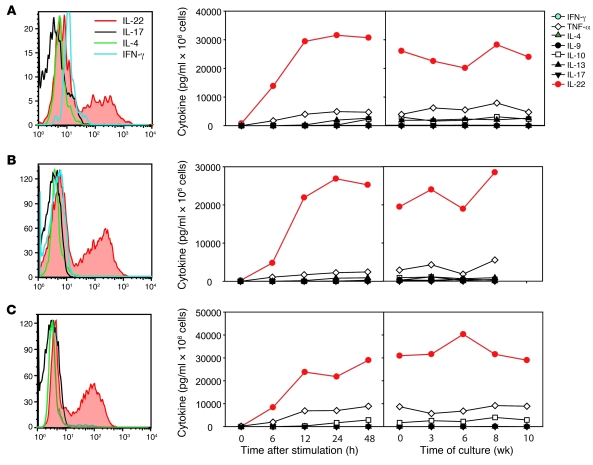
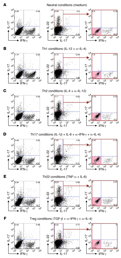
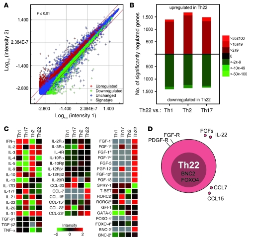
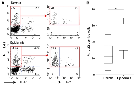
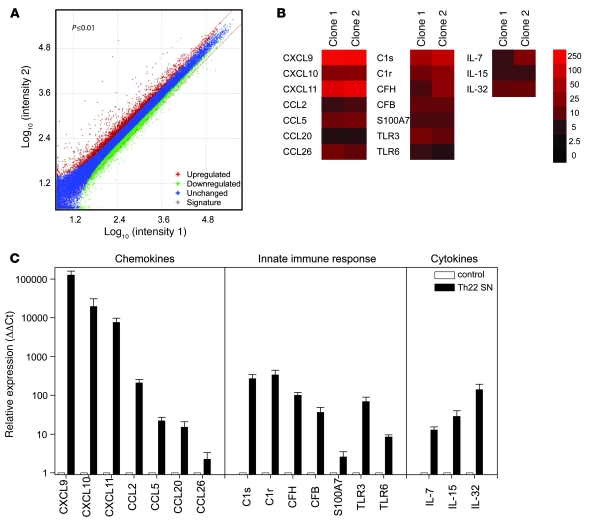
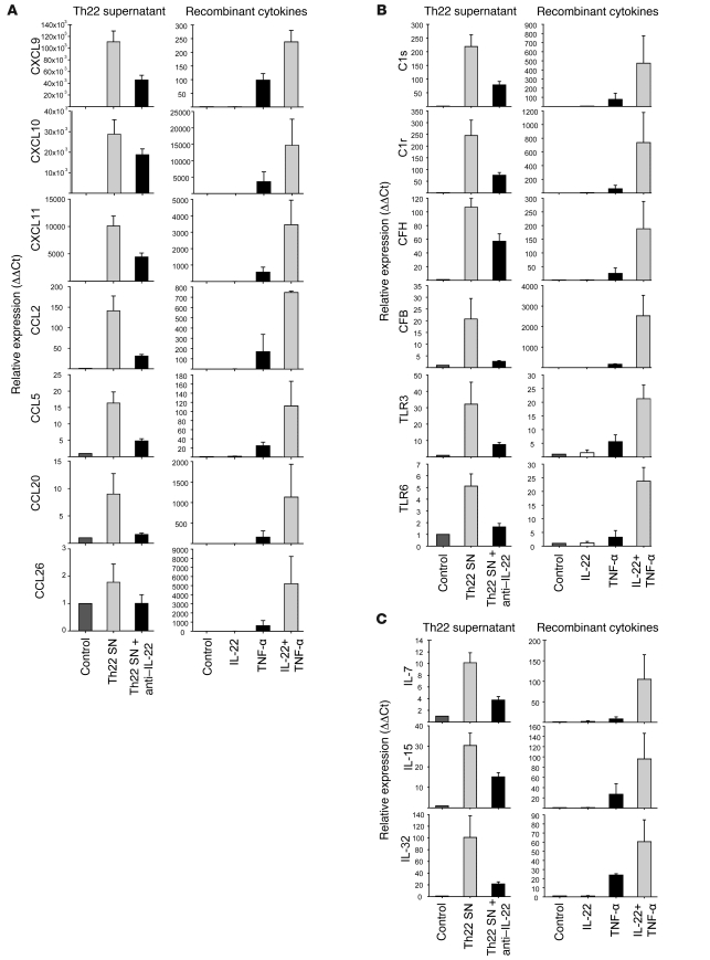
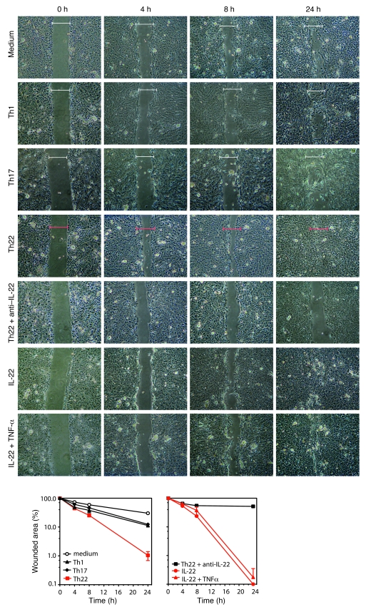
Th subsets are defined according to their production of lineage-indicating cytokines and functions. In this study, we have identified a subset of human Th cells that infiltrates the epidermis in individuals with inflammatory skin disorders and is characterized by the secretion of IL-22 and TNF-alpha, but not IFN-gamma, IL-4, or IL-17. In analogy to the Th17 subset, cells with this cytokine profile have been named the Th22 subset. Th22 clones derived from patients with psoriasis were stable in culture and exhibited a transcriptome profile clearly separate from those of Th1, Th2, and Th17 cells; it included genes encoding proteins involved in tissue remodeling, such as FGFs, and chemokines involved in angiogenesis and fibrosis. Primary human keratinocytes exposed to Th22 supernatants expressed a transcriptome response profile that included genes involved in innate immune pathways and the induction and modulation of adaptive immunity. These proinflammatory Th22 responses were synergistically dependent on IL-22 and TNF-alpha. Furthermore, Th22 supernatants enhanced wound healing in an in vitro injury model, which was exclusively dependent on IL-22. In conclusion, the human Th22 subset may represent a separate T cell subset with a distinct identity with respect to gene expression and function, present within the epidermal layer in inflammatory skin diseases. Future strategies directed against the Th22 subset may be of value in chronic inflammatory skin disorders.


Figures

Figure 1
Figure 1. Th22 cells represent a distinct T cell subset enriched in inflammatory skin diseases.
(AD) Representative 4-color intracellular cytokine stainings of primary human T lymphocytes derived from PBMCs (A), PS (B), AE (C), and ACD (D) for IL-22, IL-17, IFN-γ, and IL-4. Shown are dot plots for IL-22 and IL-17 and of IL-17IL-22+ gated cells (red outline) for IFN-γ and IL-4. Th22 cells are shown by red shading. Numbers indicate relative percentages per quadrant. (E) Frequency of IL-22+ cells in PBMCs (n = 4) and in PS (n = 3), AE (n = 4), and ACD (n = 3). Symbols denote individual determinations, and horizontal bars denote mean. *P < 0.05; **P < 0.01. (F) Plasticity in T cell phenotypes, as shown by coexpression of IL-22 with other cytokines in PS (n = 3), AE (n = 4), and ACD (n = 4). Gray ovals denote scale, indicating 1% of total cells. (G) The majority of IL-22+ T cells belongs to the CD4+ subpopulation, as shown for CD4/CD8 sorted cells from PBMCs and skin T cell lines of PS, AE, and ACD. Numbers indicate relative percentages per quadrant.
Figure 2
Figure 2. Th22 clones show a stable phenotype.
Skin T cell lines were cloned by limiting dilution and analyzed by intracellular cytokine staining and ELISA. Th22 clones 1 (A), 2 (B), and 3 (C; see Table 1) produced exclusively IL-22, as determined by multicolor flow cytometry. Stability of the Th22 phenotype is also shown in a time course after TCR stimulation and over a culture time of 10 weeks with subsequent TCR restimulations. Cytokine content of cell-free supernatant is shown for 1 representative experiment.
Figure 3
Figure 3. Th22 cells are stable and cannot be deformed into other T cell phenotypes.
Freshly isolated CCR10+ T cells did not convert into another Th subset. Shown are intracellular stainings for CCR10+CD45RA cells after 5 days under nonpolarizing (A), Th1 (B), Th2 (C), Th17 (D), Th22 (E), and Treg (F) conditions and subsequent stimulation with PMA/ionomycin. Shown are dot plots for IL-17 and IFN-γ, for IL-22 and IL-17, and of IL-17IL-22+ gated cells (red outline) for IFN-γ and IL-4. Th22 cells are shown by red shading. Numbers indicate relative percentages per quadrant. Data are representative for 3 independent donors. Mean values and corresponding IL-22, TNF-α, IL-13, and IL-4 secretion are shown in Supplemental Figure 3.
Figure 4
Figure 4. Whole genome transcriptome analysis of Th22 compared with Th1, Th2, and Th17 cells reveals unique functional profiling of Th22.
Th22 clones (n = 3) and Th1, Th2, and Th17 clones (n = 5) were stimulated for 6 hours with a TCR stimulus, and the mRNA expression profile was analyzed in a whole genome microarray approach. (A) Differential Th22 transcriptome, shown as a dLogPlot of Th22 pool genes that were upregulated or downregulated compared with Th1, Th2, and Th17 pools. (B) Th22 transcriptome separation, shown by total up- and downregulated genes in the Th22 pool compared with the single subsets of Th1, Th2, and Th17 clones. (C) Clonal phenotypes. Intensity of immunologically relevant genes of T cell subsets is shown as a heatmap. The Agilent array (single color, technology 14850) contains multiple probe sets for some genes that were empirically observed to differ in intensity from a gene-covering selection of probes. The differential abundance of these probes can originate from alternative splice variants or differential mRNA stability, for example. Alternative probes showing differences in intensity in the present study are indicated by Roman numerals; probe set sequences are available at http://www.agilent.com. Compared with CCL15I (A_23_P218369), CCL15II (A_24_P301501) recognizes an additional splice variant. For FGF1, 16 splice variants are already suggested, and the difference between Th1 and Th22 may originate in differential exon usage (shown are FGF1I, A_23_P136433; FGF1II, A_24_P251969; FGF1III, A_24_P111106; FGF1IV, A_23_P213336). For FGF12, 11 different splice variants are known, and differential signals could reflect differential splicing (shown are FGF12I, A_24_P334300; FGF12II, A_23_P211727). Probe RORC2II (A_23_P372910) binds in the 3′ untranslated region, whereas RORC2I (A_23_P324107) binds in the coding region. FOXO4II (A_24_P379165) binds the 3′ end of the gene, for which up to 3 splice variants may exist (also shown is FOXO4I, A_24_P911066). The more sensitive BNC-2I (A_23_P43690) binds exon 6, whereas BNC-2II (A_23_P43684) binds to the C-terminal exon 7. For CCL23, 2 splice variants are known, of which variant 1 is recognized by CCL23I (A_24_P319088) and both variants by CCL23II (A_24_P133905). (D) Overview of the Th22 phenotype.
Figure 5
Figure 5. Th22 cells are enriched in the epidermal compartment of the skin.
Skin biopsies were separated into dermal and epidermal parts by dispase treatment. Dermal and epidermal T cell lines were analyzed by flow cytometry. (A) Representative 3-color intracellular staining for IL-22, IL-17, and IFN-γ (on Il-17IL-22+ gated cells; red outline) of a separated ACD biopsy. Numbers indicate relative percentages in each quadrant. (B) Calculated mean percentage of total IL-22+ cells in dermis and epidermis (n = 4 each of AE and ACD). Lines within boxes denote means; box upper and lower bounds indicate SD; and whiskers indicate minimum and maximum values. *P < 0.05.
Figure 6
Figure 6. Th22 cells induce a specific response pattern in primary human keratinocytes.
Primary human keratinocytes (n = 3) were stimulated for 12 hours with supernatants (SN) of activated Th22 clones (n = 2). mRNA was extracted and hybridized to whole genome microarrays. (A) dLogPlot of up- and downregulated genes in primary human keratinocytes in response to Th22 supernatant incubation. (B) Extract of significantly upregulated genes by supernatants of Th22 clones. (C) Confirmation of array results by real-time PCR. Data are representative for at least 3 independent experiments. Error bars indicate SD.
Figure 7
Figure 7. Th22 cells influence keratinocyte functions by a combination of IL-22 and TNF-α.
Human primary keratinocytes were stimulated with Th22 supernatants in the presence or absence of neutralizing anti–IL-22 antibodies (left) or with recombinant cytokines (right). mRNA was extracted after 12 hours, and gene expression was analyzed by real-time PCR. (A) Chemokine expression. (B) Innate immune response. (C) Cytokine expression pattern. Data are representative for at least 3 independent experiments. Error bars indicate SD.
Figure 8
Figure 8. Th22 supernatant enhances wound healing in a functional in vitro injury model.
Microscopic photography after wound induction on a confluent monolayer of primary human keratinocytes in a time course from 4 to 24 hours revealed that wound healing was efficiently enhanced by Th22 supernatant and by IL-22, but less so by other T cell subset supernatants. Neutralization of IL-22 in Th22 supernatant reverted this effect. Brackets show initial wound size for comparison. Graphs below show relative wound closure over time, based on the wound gap compared with initial wound size. Error bars indicate SD.

Similar articles

Cited by

References

    1. Burgler S., et al. 2009Differentiation and functional analysis of human T(H)17 cells. J. Allergy Clin. Immunol. 123588–595; e581–e587. . 10.1016/j.jaci.2008.12.017 - DOI - PubMed
    1. Eyerich K., et al. IL-17 in atopic eczema: linking allergen-specific adaptive and microbial-triggered innate immune response. J. Allergy Clin. Immunol. 2009;123:59–66; e54. doi: 10.1016/j.jaci.2008.10.031. - DOI - PubMed
    1. Aujla S.J., et al. IL-22 mediates mucosal host defense against Gram-negative bacterial pneumonia. Nat. Med. 2008;14:275–281. doi: 10.1038/nm1710. - DOI - PMC - PubMed
    1. Schmidt-Weber C.B., Akdis M., Akdis C.A. TH17 cells in the big picture of immunology. J. Allergy Clin. Immunol. 2007;120:247–254. doi: 10.1016/j.jaci.2007.06.039. - DOI - PubMed
    1. Liang S.C., et al. Interleukin (IL)-22 and IL-17 are coexpressed by Th17 cells and cooperatively enhance expression of antimicrobial peptides. J. Exp. Med. 2006;203:2271–2279. doi: 10.1084/jem.20061308. - DOI - PMC - PubMed

Publication types

MeSH terms