. 2009 Dec;119(12):3817-29.
doi: 10.1172/JCI39054. Epub 2009 Nov 16.

MAPK phosphatase-1 facilitates the loss of oxidative myofibers associated with obesity in mice

Affiliations

MAPK phosphatase-1 facilitates the loss of oxidative myofibers associated with obesity in mice

Rachel J Roth et al. J Clin Invest. 2009 Dec.

Abstract

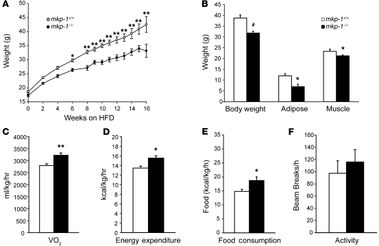
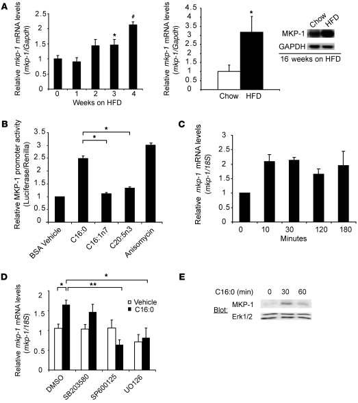
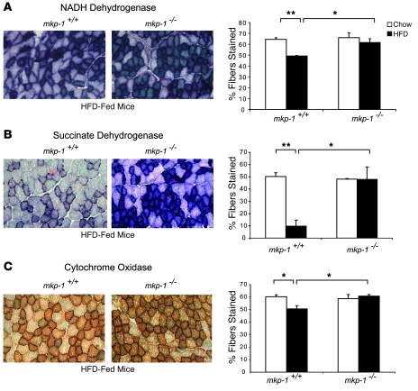
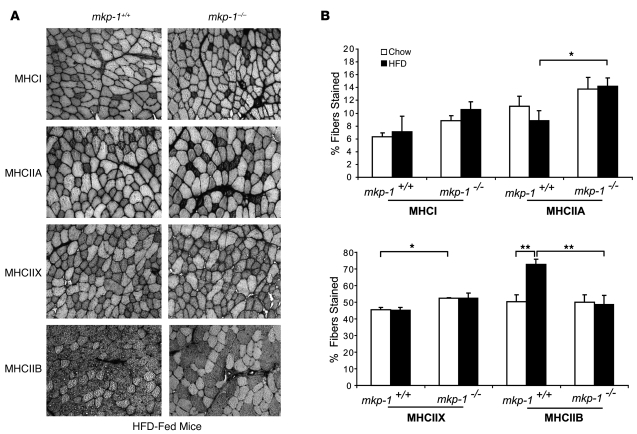
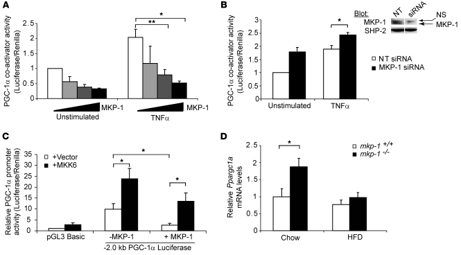
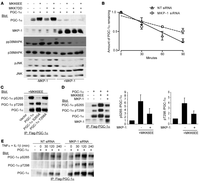
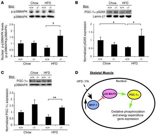
Oxidative myofibers, also known as slow-twitch myofibers, help maintain the metabolic health of mammals, and it has been proposed that decreased numbers correlate with increased risk of obesity. The transcriptional coactivator PPARgamma coactivator 1alpha (PGC-1alpha) plays a central role in maintaining levels of oxidative myofibers in skeletal muscle. Indeed, loss of PGC-1alpha expression has been linked to a reduction in the proportion of oxidative myofibers in the skeletal muscle of obese mice. MAPK phosphatase-1 (MKP-1) is encoded by mkp-1, a stress-responsive immediate-early gene that dephosphorylates MAPKs in the nucleus. Previously we showed that mice deficient in MKP-1 have enhanced energy expenditure and are resistant to diet-induced obesity. Here we show in mice that excess dietary fat induced MKP-1 overexpression in skeletal muscle, and that this resulted in reduced p38 MAPK-mediated phosphorylation of PGC-1alpha on sites that promoted its stability. Consistent with this, MKP-1-deficient mice expressed higher levels of PGC-1alpha in skeletal muscle than did wild-type mice and were refractory to the loss of oxidative myofibers when fed a high-fat diet. Collectively, these data demonstrate an essential role for MKP-1 as a regulator of the myofiber composition of skeletal muscle and suggest a potential role for MKP-1 in metabolic syndrome.


Figures

Figure 1
Figure 1. Resistance to diet-induced obesity and increased energy expenditure in mkp-1–/– mice.
(A) 129/J mkp-1+/– mice were backcrossed to wild-type C57BL6/J mice for 8 generations. At weaning, backcrossed mkp-1+/+ and mkp-1–/– mice were placed on a HFD, and weights were monitored weekly for 16 weeks. Data are mean ± SEM (n = 8–16 per time point). (B) mkp-1+/+ and mkp-1–/– mice were subjected to proton magnetic resonance spectroscopy analysis after 16 weeks of HFD feeding (n = 6–8). Data are mean ± SEM. (CF) mkp-1+/+ and mkp-1–/– mice were subjected to open circuit calorimetry after 16 weeks of HFD feeding, and (C) oxygen consumption, (D) energy expenditure, (E) food consumption, and (F) locomotor activity were recorded. Data in CF are mean ± SEM (n = 6–8). *P < 0.05, **P < 0.005, #P < 0.0005 versus mkp-1+/+.
Figure 2
Figure 2. HFD and FAs induce MKP-1 overexpression in skeletal muscle.
(A) Quadriceps were isolated from mice fed chow or HFD for the indicated times. Northern blots were performed for MKP-1 and normalized to GAPDH. Shown are normalized mkp-1 mRNA levels for 0–4 weeks of HFD (n = 3–9) or for 16-week chow- or HFD-fed mice (n = 6–7). A representative Northern blot image is shown for the latter. Data are mean ± SEM. (B) C2C12 myoblasts were transfected with MKP-1 promoter–luciferase (–1.4 kb) and TK-Renilla and stimulated for 16 hours with 500 μM palmitate (C16:0), palmitoleate (C16:1n7), eicosapentaenoic acid (C20:5n3), and anisomycin. Data are mean ± SEM of luciferase normalized to Renilla relative to vehicle control (n = 3–7). (C) C2C12 myoblasts were stimulated with 500 μM palmitate for the indicated times, RNA was harvested, and mkp-1 mRNA levels were measured by quantitative real-time RT-PCR and normalized to 18S. Data are mean ± SEM from 3–7 experiments. (D) C2C12 myoblasts were pretreated with the indicated MAPK inhibitors and stimulated with vehicle or 500 μM palmitate for 30 minutes. MKP-1 levels were determined as in C. Data are mean ± SEM from 3–7 experiments. (E) C2C12 myoblasts were stimulated for the indicated times with 500 μM palmitate and immunoblotted for MKP-1 or Erk1/2. Immunoblot is representative of 3 separate experiments. *P < 0.05, **P < 0.005, #P < 0.0005 versus respective control or as otherwise indicated by brackets.
Figure 3
Figure 3. MKP-1 mediates oxidative myofiber loss in obesity.
(A) NADH dehydrogenase stain and quantitation (n = 5–6) of TA muscle from HFD-fed mkp-1+/+ and mkp-1–/– mice. (B) Succinate dehydrogenase stain and quantitation (n = 3) of TA muscle from HFD-fed mkp-1+/+ and mkp-1–/– mice. (C) Cytochrome oxidase stain and quantitation (n = 3–5) of TA muscle from HFD-fed mkp-1+/+ and mkp-1–/– mice. Original magnification, ×100. Data are mean ± SEM. *P < 0.05; **P < 0.005.
Figure 4
Figure 4. MKP-1 mediates a glycolytic fiber type switch in obesity.
(A) Photomicrographs representing MHCI, MHCIIA, MHCIIX, and MHCIIB immunohistochemical staining of TA muscle from HFD-fed mkp-1+/+ and mkp-1–/– mice. Original magnification, ×100. (B) Percentage of fiber type stained (n = 4–8). Data are mean ± SEM. *P < 0.05; **P < 0.005.
Figure 5
Figure 5. MKP-1 negatively regulates PGC-1α coactivator activity.
(A) PGC-1α–GAL4, 5xGAL4-luciferase, increasing concentrations of MKP-1, and TK-Renilla were transfected into C2C12 myoblasts. Myoblasts were stimulated with 10 ng/ml TNF-α for 24 hours prior to measurement of luciferase and Renilla. Data are mean ± SEM normalized luciferase/Renilla values from 4 separate experiments. (B) Left: PGC-1α–GAL4, 5xGAL4-luciferase, and TK-Renilla were transfected into C2C12 myoblasts along with 50 nM MKP-1 or nontargeting (NT) siRNA. Myoblasts were starved and either left unstimulated or stimulated with 10 ng/ml TNF-α for 24 hours prior to measurement of luciferase and Renilla. Data are mean ± SEM normalized luciferase/Renilla from 3 separate experiments. Right: C2C12 myoblasts were transfected with 50 nM of either MKP-1 or nontargeting siRNA; 24 hours later, cells were immunoblotted for MKP-1 or SHP-2 as a loading control. Lanes were run on the same gel but were noncontiguous (white line). (C) pGL3-PGC-1α promoter–luciferase (–2.0 kb) and TK-Renilla were transfected into C2C12 myoblasts in the absence or presence of MKP-1 and MKK6EE. Luciferase and Renilla were measured 48 hours later. Data are mean ± SEM normalized luciferase/Renilla values from 4 separate experiments. (D) Ppargc1a mRNA levels in TA muscle of age-matched mkp-1+/+ and mkp-1–/– mice after 16 weeks of chow or HFD, as measured by quantitative real-time RT-PCR. Data are mean ± SEM normalized to 18S mRNA (n = 5–8). *P < 0.05; **P < 0.005.
Figure 6
Figure 6. MKP-1 antagonizes PGC-1α phosphorylation and protein stability.
(A) Flag-tagged PGC-1α, MKK6EE, MKK7DD, and MKP-1 (10 μg) were transfected into C2C12 myoblasts. Myoblasts were lysed 24 hours later and immunoblotted with PGC-1α, MKP-1, phospho-p38 MAPK, p38 MAPK, phospho-JNK, and JNK antibodies. (B) Flag-tagged PGC-1α was transfected into C2C12 myoblasts along with nontargeting siRNA or MKP-1 siRNA as described in Figure 5B, and pulse-chase metabolic labeling with 35S methionine/cysteine was performed. Densitometric analyses were quantitated as the amount of PGC-1α remaining relative to time 0. The mean t1/2 was calculated from the linear regression derived from 3 independent experiments (nontargeting siRNA, 56.7 minutes; MKP-1 siRNA, 83.5 minutes; P < 0.05). Data are mean ± SEM. (C) Flag-tagged PGC-1α as well as Flag-tagged PGC-1α Ser265A and Thr298A mutants were transfected into C2C12 myoblasts with MKK6EE. Myoblasts were lysed, and PGC-1α was immunoprecipitated with anti-Flag antibodies and immunoblotted with total PGC-1α, phospho-Ser265 PGC-1α, or phospho-Thr298 PGC-1α antibodies. Lanes were run on the same gel but were noncontiguous (white line). (D) Flag-PGC-1α, MKK6EE, and MKP-1 (2 μg) were transfected into C2C12 myoblasts. Myoblasts were lysed, and PGC-1α was immunoprecipitated with anti-Flag antibodies and immunoblotted as in C. Graphs show densitometric measurements of phospho-Ser265 PGC-1α and phospho-Thr298 PGC-1α normalized to total PGC-1α from 3 independent experiments. Data are mean ± SEM. (E) Flag-tagged PGC-1α was transfected into C2C12 myoblasts along with 50 nM nontargeting or MKP-1 siRNA. Myoblasts were starved for 2 hours prior to stimulation with 10 ng/ml TNF-α and 2 ng/ml IL-1β for the indicated times. Myoblasts were lysed, and PGC-1α was immunoprecipitated with anti-Flag antibodies and immunoblotted as in C.
Figure 7
Figure 7. Enhanced PGC-1α expression and phosphorylation in mkp-1–/– mice.
(A) Nuclear extracts of TA muscle from age-matched mkp-1+/+ and mkp-1–/– mice fed chow or HFD for 16 weeks were immunoblotted for phospho–p38 MAPK or p38 MAPK. Also shown are densitometric measurements for phospho–p38 MAPK levels normalized to total p38 MAPK. Data are mean ± SEM (n = 8–9). (B) Nuclear extracts as in A were immunoblotted for phospho-Ser265 PGC-1α. Lanes were run on the same gel but were noncontiguous (white line). Also shown are densitometric measurements for phospho-Ser265 PGC-1α expression normalized to Lamin-β1. Data are mean ± SEM (n = 4–9). (C) TA muscle was isolated from mkp-1+/+ and mkp-1–/– mice fed chow or HFD for 16 weeks. Muscle lysates were immunoblotted with PGC-1α antibodies, and densitometric measurements for PGC-1α expression were normalized to p38 MAPK. Data are mean ± SEM (n = 6). (D) Under HFD conditions, exposure to FAs increases MKP-1 expression, causing inactivation of nuclear p38 MAPK. Reduced p38 MAPK–mediated phosphorylation of PGC-1α decreases PGC-1α expression. Impaired skeletal muscle PGC-1α function facilitates loss of oxidative myofiber composition. P = 0.05; *P < 0.05; **P < 0.005.

Similar articles

Cited by

References

    1. Raman M., Chen W., Cobb M.H. Differential regulation and properties of MAPKs. Oncogene. 2007;26:3100–3112. doi: 10.1038/sj.onc.1210392. - DOI - PubMed
    1. Weston C.R., Davis R.J. The JNK signal transduction pathway. Curr. Opin. Cell Biol. 2007;19:142–149. doi: 10.1016/j.ceb.2007.02.001. - DOI - PubMed
    1. Cuenda A., Rousseau S. p38 MAP-Kinases pathway regulation, function and role in human diseases. Biochim. Biophys. Acta. . 2007;1773:1358–1375. doi: 10.1016/j.bbamcr.2007.03.010. - DOI - PubMed
    1. Murphy L.O., Blenis J. MAPK signal specificity: the right place at the right time. Trends Biochem. Sci. 2006;31:268–275. doi: 10.1016/j.tibs.2006.03.009. - DOI - PubMed
    1. Weston C.R., Davis R.J. The JNK signal transduction pathway. Curr. Opin. Cell Biol. 2007;19:142–149. doi: 10.1016/j.ceb.2007.02.001. - DOI - PubMed

Publication types

MeSH terms