

A PI3 kinase inhibitor found to activate bestrophin 3
- PMID: 19920768
- PMCID: PMC3052421
- DOI: 10.1097/FJC.0b013e3181c87c85
A PI3 kinase inhibitor found to activate bestrophin 3
Abstract
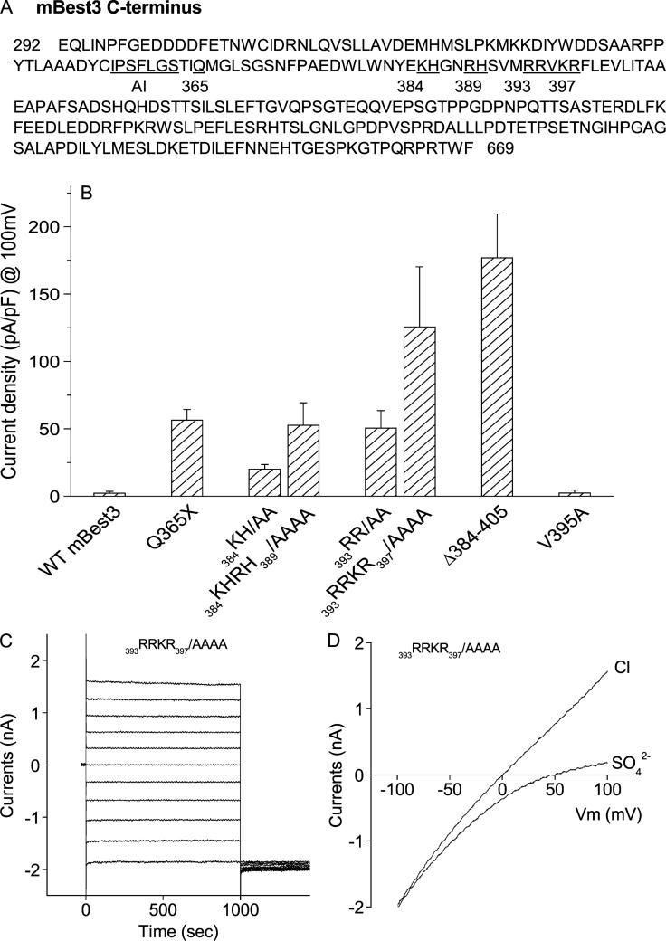
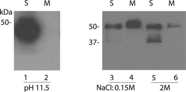
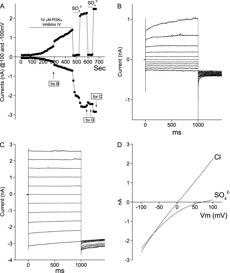
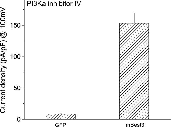
Bestrophin 3 (Best3), a member of bestrophin Cl channel family, is a CaCl(cGMP) channel candidate in vascular smooth muscle cells. The mechanism for its activation remains unclear. In previous studies, we reported that an autoinhibitory domain ((356)IPSFLGS(362)) existed in Best3 C-terminus and when the autoinhibitory domain was mutated, the Best3 channel was dramatically activated. In this study, we further dissected the roles of the C-terminal sequence in Best3 activation. We found that there were eight basic amino acids downstream of the AI domain within the region (384-397), which were also involved in Best3 activation. Mutations of these basic amino acids significantly activated Best3 as a Cl channel. Led by the assumption that the basic amino acids may be involved in the Best3 C-terminal membrane association through binding to membranous phospholipids, we discovered that PI3Kalpha inhibitor IV could strongly activate Best3.
Figures
Similar articles
-
The bestrophin- and TMEM16A-associated Ca(2+)- activated Cl(–) channels in vascular smooth muscles.Channels (Austin). 2014;8(4):361-9. doi: 10.4161/chan.29531. Channels (Austin). 2014. PMID: 25478625 Free PMC article. Review.
-
C-terminal membrane association of Bestrophin 3 and its activation as a chloride channel.J Membr Biol. 2013 Feb;246(2):177-82. doi: 10.1007/s00232-012-9514-7. Epub 2012 Nov 3. J Membr Biol. 2013. PMID: 23124946
-
Activation of bestrophin Cl- channels is regulated by C-terminal domains.J Biol Chem. 2007 Jun 15;282(24):17460-7. doi: 10.1074/jbc.M701043200. Epub 2007 Apr 17. J Biol Chem. 2007. PMID: 17442670
-
A variant of the Ca2+-activated Cl channel Best3 is expressed in mouse exocrine glands.J Membr Biol. 2008 Mar;222(1):43-54. doi: 10.1007/s00232-008-9098-4. J Membr Biol. 2008. PMID: 18414923
-
Mechanisms of cellular synchronization in the vascular wall. Mechanisms of vasomotion.Dan Med Bull. 2010 Oct;57(10):B4191. Dan Med Bull. 2010. PMID: 21040688 Review.
Cited by
-
The bestrophin- and TMEM16A-associated Ca(2+)- activated Cl(–) channels in vascular smooth muscles.Channels (Austin). 2014;8(4):361-9. doi: 10.4161/chan.29531. Channels (Austin). 2014. PMID: 25478625 Free PMC article. Review.
-
Bestrophin-encoded Ca²⁺-activated Cl⁻ channels underlie a current with properties similar to the native current in the moth Spodoptera littoralis olfactory receptor neurons.PLoS One. 2012;7(12):e52691. doi: 10.1371/journal.pone.0052691. Epub 2012 Dec 26. PLoS One. 2012. PMID: 23300744 Free PMC article.
-
C-terminal membrane association of Bestrophin 3 and its activation as a chloride channel.J Membr Biol. 2013 Feb;246(2):177-82. doi: 10.1007/s00232-012-9514-7. Epub 2012 Nov 3. J Membr Biol. 2013. PMID: 23124946
References
-
- Petrukhin K, Koisti MJ, Bakall B, et al. Identification of the gene responsible for Best macular dystrophy. Nat Genet. 1998;19:241–247. - PubMed
-
- Marquardt A, Stohr H, Passmore LA, et al. Mutations in a novel gene, VMD2, encoding a protein of unknown properties cause juvenile-onset vitelliform macular dystrophy (Best's disease). Hum Mol Genet. 1998;7:1517–1525. - PubMed
-
- Hartzell C, Qu Z, Putzier I, et al. Looking chloride channels straight in the eye: bestrophins, lipofuscinosis, and retinal degeneration. Physiology. 2005;20:292–302. - PubMed
-
- Hartzell HC, Qu Z, Yu K, et al. Molecular physiology of bestrophins: multifunctional membrane proteins linked to best disease and other retinopathies. Physiol Rev. 2008;88:639–672. - PubMed
Publication types
MeSH terms
Substances
Grants and funding
LinkOut - more resources
Full Text Sources
