. 2009 Dec 15;101(12):2005-14.
doi: 10.1038/sj.bjc.6605437. Epub 2009 Nov 17.

Glycogen synthase kinase-3: a new therapeutic target in renal cell carcinoma

Affiliations

Glycogen synthase kinase-3: a new therapeutic target in renal cell carcinoma

V Bilim et al. Br J Cancer. .

Abstract

Background: Renal cell carcinoma (RCC) is highly resistant to chemotherapy because of a high apoptotic threshold. Recent evidences suggest that GSK-3beta positively regulates human pancreatic cancer and leukaemia cell survival in part through regulation of nuclear factor (NF-kappaB)-mediated expression of anti-apoptotic molecules. Our objectives were to determine the expression pattern of GSK-3beta and to assess the anti-cancer effect of GSK-3beta inhibition in RCC.

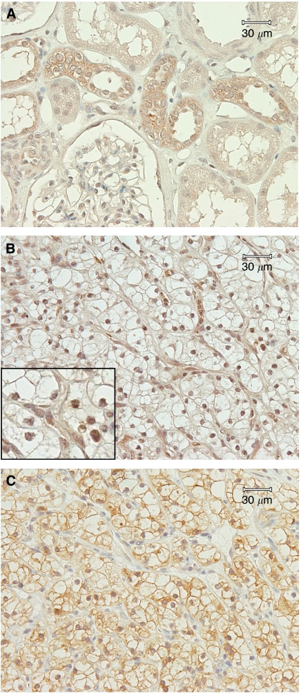
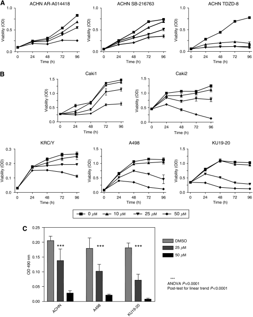
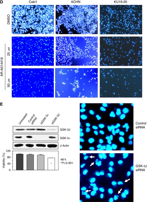
Methods: Immunohistochemistry and nuclear/cytosolic fractionation were performed to determine the expression pattern of GSK-3beta in human RCCs. We used small molecule inhibitor, RNA interference, western blotting, quantitative RT-PCR, BrDU incorporation and MTS assays to study the effect of GSK-3beta inactivation on renal cancer cell proliferation and survival.

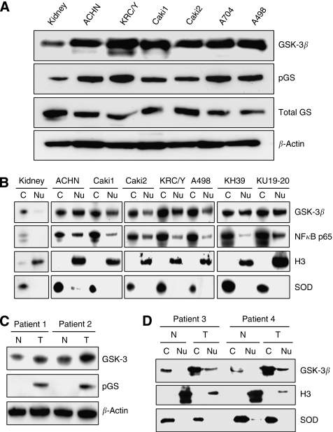
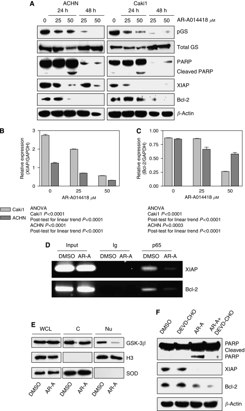
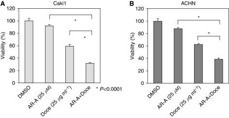
Results: We detected aberrant nuclear accumulation of GSK-3beta in RCC cell lines and in 68 out of 74 (91.89%) human RCCs. We found that pharmacological inhibition of GSK-3 led to a decrease in proliferation and survival of renal cancer cells. We observed that inhibition of GSK-3 results in decreased expression of NF-kappaB target genes Bcl-2 and XIAP and a subsequent increase in renal cancer cell apoptosis. Moreover, we show that GSK-3 inhibitor and Docetaxel synergistically suppress proliferation and survival of renal cancer cells.

Conclusions: Our results show nuclear accumulation of GSK-3beta as a new marker of human RCC, identify that GSK-3 positively regulates RCC cell survival and proliferation and suggest inhibition of GSK-3 as a new promising approach in the treatment of human renal cancer.


Figures

Figure 1
Figure 1
GSK-3β is overexpressed in nuclei of renal cancer cells. (A) Protein lysates from the indicated RCC cell lines and normal kidney as a control were separated by SDS–PAGE (50 μg per well), transferred to PVDF membrane and probed with antibodies against GSK-3β, phospho-glycogen synthase (pGS) and total glycogen synthase (GS). (B) Cytosolic (C) and nuclear (Nu) fractions were prepared from RCC cell lines and normal kidney, separated by SDS–PAGE (50 μg per well), transferred to PVDF membrane and probed with indicated antibodies. Cu/Zn supeoxide dismutase (SOD) and histone H3 (H3) were used as cytosolic and nuclear markers, respectively. (C) Expression of GSK-3β and pGS was detected in protein extracts from primary tumour (T) and corresponding normal kidney tissue (N) obtained from kidney cancer patients. (D) Nuclear (Nu) and cytosolic (C) fractions were prepared from fresh tumour (T) and corresponding normal kidney tissue (N) sampled from kidney cancer patients, and analysed as described in (B).
Figure 2
Figure 2
Immunohistochemical analysis of GSK-3β expression in normal human kidney (A). Immunohistochemical analysis of GSK-3β (B) and pGS (C) expression in serial sections of renal carcinoma. Insert in (B) shows higher magnification view.
Figure 3
Figure 3
Inhibition of GSK-3 suppresses proliferation of renal cancer cells. (A) Relative cell viability was measured by MTS assay in ACHN renal cancer cell line treated with indicated doses of AR-A014418, SB-216763 or TDZD-8 for 24, 48, 72 and 96 h. (B) Relative cell viability was measured by MTS assay in RCC cell lines treated with indicated doses of AR-A014418 for 24, 48, 72 and 96 h. (C) ACHN, A498 and KU19-20 renal cancer cells were treated with diluent (DMSO) or AR-A014418 with indicated doses for 48 h. BrdU colometric assay was performed as described in ‘Materials and Methods’. The results are presented as OD 490 nm (ANOVA P<0.0001, post test for linear trend P<0.0001). (D) ACHN, Caki1 and KU19-20 renal cancer cells were cultured in the presence of DMSO or indicated concentrations of AR-A014418 for 96 h, followed by Hoechst 33342 staining. (E) ACHN renal cancer cells were transfected with control siRNA, GSK-3β or GSK-3α siRNA using Lipofectamine; 48 h after transfection, relative cell viability was measured in transfected cancer cells by MTS assay as shown in lower panel. Western blot for GSK-3α, GSK-3β and actin as control for loading is presented in the upper panel. Right panel represents Hoechst 33342 staining of ACHN cells transfected with control siRNA (right-upper) or GSK-3β siRNA (right-lower). Apoptotic cells are indicated by arrows.
Figure 3
Figure 3
Inhibition of GSK-3 suppresses proliferation of renal cancer cells. (A) Relative cell viability was measured by MTS assay in ACHN renal cancer cell line treated with indicated doses of AR-A014418, SB-216763 or TDZD-8 for 24, 48, 72 and 96 h. (B) Relative cell viability was measured by MTS assay in RCC cell lines treated with indicated doses of AR-A014418 for 24, 48, 72 and 96 h. (C) ACHN, A498 and KU19-20 renal cancer cells were treated with diluent (DMSO) or AR-A014418 with indicated doses for 48 h. BrdU colometric assay was performed as described in ‘Materials and Methods’. The results are presented as OD 490 nm (ANOVA P<0.0001, post test for linear trend P<0.0001). (D) ACHN, Caki1 and KU19-20 renal cancer cells were cultured in the presence of DMSO or indicated concentrations of AR-A014418 for 96 h, followed by Hoechst 33342 staining. (E) ACHN renal cancer cells were transfected with control siRNA, GSK-3β or GSK-3α siRNA using Lipofectamine; 48 h after transfection, relative cell viability was measured in transfected cancer cells by MTS assay as shown in lower panel. Western blot for GSK-3α, GSK-3β and actin as control for loading is presented in the upper panel. Right panel represents Hoechst 33342 staining of ACHN cells transfected with control siRNA (right-upper) or GSK-3β siRNA (right-lower). Apoptotic cells are indicated by arrows.
Figure 4
Figure 4
Inhibition of GSK-3 decreases expression of anti-apoptotic XIAP and Bcl-2 and induces apoptosis in renal cancer cells. (A) ACHN and Caki1 renal cancer cells were treated with 25 or 50 μM of AR-A014418; 24 and 48 h after treatment, the cell pellet was collected, cell lysates were separated by SDS–PAGE (50 μg per well), transferred to PVDF membrane and probed with indicated antibodies. pGS, phospho-glycogen synthase. (B, C) ACHN and Caki1 renal cancer cells were treated with 25 or 50 μM of AR-A014418; 24 h after treatment, the cell pellet was collected and RNA was extracted. Relative expression (target gene value normalised by GAPDH) of XIAP (B) and Bcl-2 (C) genes was measured by real-time PCR using TaqMan probe technique as described in ‘Materials and Methods’. P-values of ANOVA and post test for linear trend are indicated. (D) Using chromatin immunoprecipitation (ChIP) assay, binding of NF-κB p65 to the promoters of its target genes XIAP and Bcl-2 was evaluated with in ACHN RCC cells treated with DMSO or 50 μM AR-A014418 (AR-A) for 48 h. (E) ACHN renal cancer cells were treated with either DMSO or AR-A014418 (50 μmol l−1) for 24 h, nuclear/cytosolic fractions were prepared and cytozolic/nuclear GSK-3β protein was analysed as described in Figure 1B and D. (F) A498 renal cancer cells were treated with diluent (DMSO), DEVD-CHO (caspase inhibitor), AR-A014418 (AR-A) or AR-A014418+DEVD-CHO; 24 h after treatment, the cell pellet was collected and protein expression analysis was performed as described in (A).
Figure 5
Figure 5
GSK-3 inhibitor and Docetaxel synergistically suppress viability of renal cancer cells. Relative cell viability was measured by MTS assay in Caki1 (A) and ACHN (B) renal cancer cells treated with 25 μM of AR-A014418, 25 μg ml−1 Docetaxel or a combination of both for 24 h. Combined treatment with AR-014418 and Docetaxel significantly suppressed cancer cell viability (P<0.0001) compared with both agents, and the effect of combination of the two drugs was synergistic.

References

    1. An J, Sun Y, Fisher M, Rettig MB (2004) Maximal apoptosis of renal cell carcinoma by the proteasome inhibitor bortezomib is nuclear factor-kappaB dependent. Mol Cancer Ther 3: 727–736 - PubMed
    1. Beck SDW, Patel MI, Snyder ME, Kattan MW, Motzer RJ, Reuter VE, Russo P (2004) Effect of papillary and chromophobe cell type on disease-free survival after nephrectomy for renal cell carcinoma. Ann Surg Oncol 11: 71–77 - PubMed
    1. Bhat R, Xue Y, Berg S, Hellberg S, Ormo M, Nilsson Y, Radesater A-C, Jerning E, Markgren P-O, Borgegard T, Nylof M, Gimenez-Cassina A, Hernandez F, Lucas JJ, Diaz-Nido J, Avila J (2003) Structural insights and biological effects of glycogen synthase kinase 3-specific inhibitor AR-A014418. J Biol Chem 278: 45937–45945 - PubMed
    1. Bilim V, Kawasaki T, Katagiri A, Wakatsuki S, Takahashi K, Tomita Y (2000) Altered expression of beta-catenin in renal cell cancer and transitional cell cancer with the absence of beta-catenin gene mutations. Clin Cancer Res 6: 460–466 - PubMed
    1. Bilim V, Yuuki K, Itoi T, Muto A, Kato T, Nagaoka A, Motoyama T, Tomita Y (2008) Double inhibition of XIAP and Bcl-2 axis is beneficial for retrieving sensitivity of renal cell cancer to apoptosis. Br J Cancer 98: 941–949 - PMC - PubMed

Publication types

MeSH terms