

Longitudinal in vivo imaging reveals balanced and branch-specific remodeling of mature cortical pyramidal dendritic arbors after stroke
- PMID: 19920846
- PMCID: PMC2949167
- DOI: 10.1038/jcbfm.2009.241
Longitudinal in vivo imaging reveals balanced and branch-specific remodeling of mature cortical pyramidal dendritic arbors after stroke
Abstract
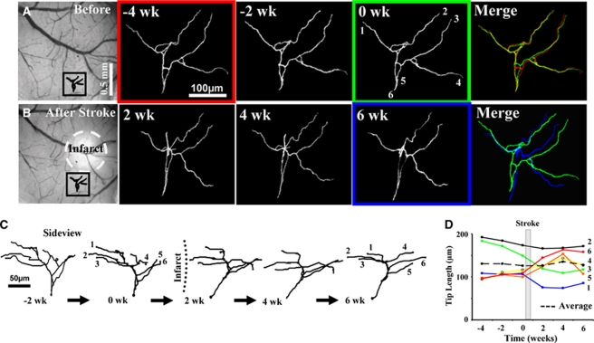
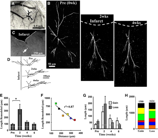
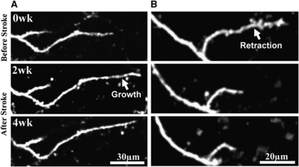
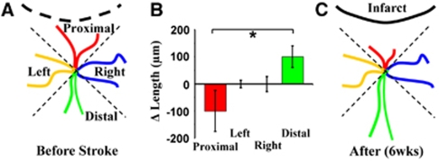
The manner in which fully mature peri-infarct cortical dendritic arbors remodel after stroke, and thus may possibly contribute to stroke-induced changes in cortical receptive fields, is unknown. In this study, we used longitudinal in vivo two-photon imaging to investigate the extent to which brain ischemia can trigger dendritic remodeling of pyramidal neurons in the adult mouse somatosensory cortex, and to determine the nature by which remodeling proceeds over time and space. Before the induction of stroke, dendritic arbors were relatively stable over several weeks. However, after stroke, apical dendritic arbor remodeling increased significantly (dendritic tip growth and retraction), particularly within the first 2 weeks after stroke. Despite a threefold increase in structural remodeling, the net length of arbors did not change significantly over time because dendrite extensions away from the stroke were balanced by the shortening of tips near the infarct. Therefore, fully mature cortical pyramidal neurons retain the capacity for extensive structural plasticity and remodel in a balanced and branch-specific manner.
Figures
Similar articles
-
Absence of large-scale dendritic plasticity of layer 5 pyramidal neurons in peri-infarct cortex.J Neurosci. 2011 Feb 2;31(5):1734-8. doi: 10.1523/JNEUROSCI.4386-10.2011. J Neurosci. 2011. PMID: 21289182 Free PMC article.
-
Extensive turnover of dendritic spines and vascular remodeling in cortical tissues recovering from stroke.J Neurosci. 2007 Apr 11;27(15):4101-9. doi: 10.1523/JNEUROSCI.4295-06.2007. J Neurosci. 2007. PMID: 17428988 Free PMC article.
-
Chronic in vivo imaging shows no evidence of dendritic plasticity or functional remapping in the contralesional cortex after stroke.Cereb Cortex. 2013 Apr;23(4):751-62. doi: 10.1093/cercor/bhs092. Epub 2012 Apr 11. Cereb Cortex. 2013. PMID: 22499800 Free PMC article.
-
Molecular control of cortical dendrite development.Annu Rev Neurosci. 2002;25:127-49. doi: 10.1146/annurev.neuro.25.112701.142932. Epub 2002 Mar 19. Annu Rev Neurosci. 2002. PMID: 12052906 Review.
-
Livin' on the edge: imaging dendritic spine turnover in the peri-infarct zone during ischemic stroke and recovery.Neuroscientist. 2008 Apr;14(2):139-46. doi: 10.1177/1073858407309854. Epub 2007 Nov 26. Neuroscientist. 2008. PMID: 18039977 Review.
Cited by
-
Factors Involved in the Functional Motor Recovery of Rats with Cortical Ablation after GH and Rehabilitation Treatment: Cortical Cell Proliferation and Nestin and Actin Expression in the Striatum and Thalamus.Int J Mol Sci. 2019 Nov 16;20(22):5770. doi: 10.3390/ijms20225770. Int J Mol Sci. 2019. PMID: 31744113 Free PMC article.
-
Macro-connectomics and microstructure predict dynamic plasticity patterns in the non-human primate brain.Elife. 2018 Nov 21;7:e34354. doi: 10.7554/eLife.34354. Elife. 2018. PMID: 30462609 Free PMC article.
-
Robust neuroprosthetic control from the stroke perilesional cortex.J Neurosci. 2015 Jun 3;35(22):8653-61. doi: 10.1523/JNEUROSCI.5007-14.2015. J Neurosci. 2015. PMID: 26041930 Free PMC article.
-
Transient global cerebral ischemia induces rapid and sustained reorganization of synaptic structures.J Cereb Blood Flow Metab. 2017 Aug;37(8):2756-2767. doi: 10.1177/0271678X16674736. Epub 2016 Jan 1. J Cereb Blood Flow Metab. 2017. PMID: 27798269 Free PMC article.
-
Excitable Adult-Generated GABAergic Neurons Acquire Functional Innervation in the Cortex after Stroke.Stem Cell Reports. 2018 Dec 11;11(6):1327-1336. doi: 10.1016/j.stemcr.2018.10.011. Epub 2018 Nov 8. Stem Cell Reports. 2018. PMID: 30416050 Free PMC article.
References
-
- Brown CE, Aminoltejari K, Erb H, Winship IR, Murphy TH. In vivo voltage-sensitive dye imaging in adult mice reveals that somatosensory maps lost to stroke are replaced over weeks by new structural and functional circuits with prolonged modes of activation within both the peri-infarct zone and distant sites. J Neurosci. 2009;29:1719–1734. - PMC - PubMed
-
- Brown CE, Wong C, Murphy TH. Rapid morphologic plasticity of peri-infarct dendritic spines after focal ischemic stroke. Stroke. 2008;39:1286–1291. - PubMed
-
- Buonomano DV, Merzenich MM. Cortical plasticity: from synapses to maps. Annu Rev Neurosci. 1998;21:149–186. - PubMed
Publication types
MeSH terms
Grants and funding
LinkOut - more resources
Full Text Sources
Medical
