Review
. 2009 Sep-Oct;1(5):492-501.
doi: 10.1002/wnan.35.

Fluorine-containing nanoemulsions for MRI cell tracking

Affiliations
Review

Fluorine-containing nanoemulsions for MRI cell tracking

Jelena M Janjic et al. Wiley Interdiscip Rev Nanomed Nanobiotechnol. 2009 Sep-Oct.

Abstract

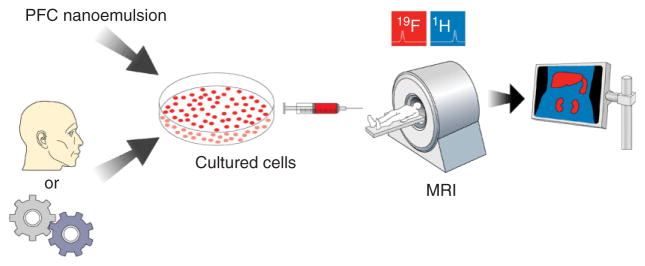
In this article we review the chemistry and nanoemulsion formulation of perfluorocarbons used for in vivo(19)F MRI cell tracking. In this application, cells of interest are labeled in culture using a perfluorocarbon nanoemulsion. Labeled cells are introduced into a subject and tracked using (19)F MRI or NMR spectroscopy. In the same imaging session, a high-resolution, conventional ((1)H) image can be used to place the (19)F-labeled cells into anatomical context. Perfluorocarbon-based (19)F cell tracking is a useful technology because of the high specificity for labeled cells, ability to quantify cell accumulations, and biocompatibility. This technology can be widely applied to studies of inflammation, cellular regenerative medicine, and immunotherapy.


Figures

FIGURE 1
FIGURE 1
Cellular MRI using PFC nanoemulsion technology. PFC nanoemulsion is added to cultured cells that have been harvested from a subject or an engineered line. The labeled cells are transplanted into the subject and imaged using 19F and 1H MRI in the same imaging session. The registered images are overlaid, yielding an image of the labeled cells in their anatomical context.
FIGURE 2
FIGURE 2
Survey of PFCs for potential MRI applications.,,–
FIGURE 3
FIGURE 3
DLS analysis of a linear PFPE nanoemulsion emulsified with F68 and PEI. Panel A displays the diameter distribution by light intensity, and B shows the diameter and PDI (error bars) followed over time at three different storage temperatures: 4 (■), 25 (●), and 37 (▼) °C. (Adapted from Ref 34).
FIGURE 4
FIGURE 4
In vivo MRI using PFC with dendritic cells (DCs) in mouse. Images of the labeled cells (i.e., 19F images) are displayed on a ‘hot-iron’ intensity scale, and the anatomical (1H) images are shown in grayscale. The three panels on the far left are a mouse quadriceps after intramuscular injection of DCs (asterisk indicates injection site). (a) 19F and 1H images (from left to right) and a ‘composite’ 19F/1H image. (b) The composite image of DC migration into the popliteal lymph node following a hind foot pad injection. (c) Composite image through the torso following intravenous inoculation with labeled DCs. Cells are apparent in the liver, spleen, and weakly in the lungs. (Data taken from Ref 19).
FIGURE 5
FIGURE 5
In vivo MRI of labeled T cells in the mouse model. The 19F image (pseudo-color) shows a localized accumulation of T cells labeled with PFPE nanoemulsion in lymph nodes and the grayscale underlay is an anatomical 1H image. Panels (a) and (b) display two consecutive 2 mm thick slices through the torso, and for anatomical orientation the kidneys (K) and gut (G) are noted. During imaging, the mouse was anesthetized with a ketamine/xylazine cocktail, connected to a mechanical ventilation apparatus, acquisitions were cardio-respiratory gated, and body temperature was regulated at 37°C. Data were collected for both 19F and 1H in a single imaging session. (Adapted from Ref 34).
FIGURE 6
FIGURE 6
Dual-mode reagents for 19F MRI and fluorescent detection. Confocal microscopy of (a) labeled mouse DCs (b) and primary T cells labeled with FBPA (red). (Adapted from Ref 34).

References

    1. Brittberg M, Lindahl A, Nilsson A, Ohlsson C, Isaksson O, et al. Treatment of deep cartilage defects in the knee with autologous chondrocyte transplantation. N Engl J Med. 1994;331(14):889–895. - PubMed
    1. Peterson L, Minas T, Brittberg M, Nilsson A, Sjogren-Jansson E, et al. Two- to 9-year outcome after autologous chondrocyte transplantation of the knee. Clin Orthop Relat R. 2000;374:212–223. - PubMed
    1. Bjorklund A, Lindvall O. Cell replacement therapies for central nervous system disorders. Nat Neurosci. 2000;6(3):537–544. - PubMed
    1. Ashley DM, Faiola B, Nair S, Hale LP, Bigner DD, et al. Bone marrow-generated dendritic cells pulsed with tumor extracts or tumor RNA induce antitumor immunity against central nervous system tumors. J Exp Med. 1997;186(7):1177–1182. - PMC - PubMed
    1. Gilboa E, Nair SK, Lyerly HK. Immunotherapy of cancer with dendritic-cell-based vaccines. Cancer Immunol Immun. 1998;46(2):82–87. - PMC - PubMed