. 2009 May 7:3:37-51.
doi: 10.2174/1874357900903010037.

Oncoviruses and Pathogenic MicroRNAs in Humans

Affiliations

Oncoviruses and Pathogenic MicroRNAs in Humans

Yoichi Robertus Fujii. Open Virol J. .

Abstract

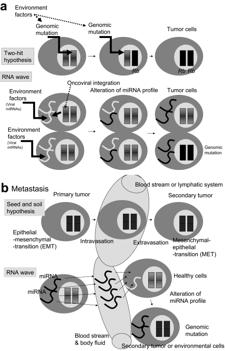
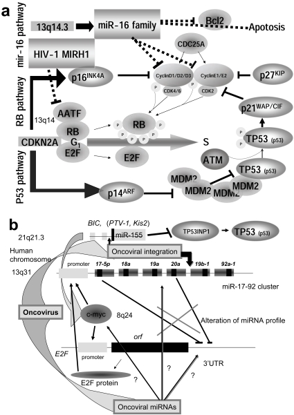
For disease prognosis, the functional significance of the oncoviral integration locus in oncogenesis has remained enigmatic. The locus encodes several transcripts without protein products, but microRNAs (miRNAs) have recently been identified from a common oncoviral integration locus. miRNA is an endogenous, non-coding small RNA by which gene expression is suppressed. Although miRNA genes, such as let-7 in the nematode, have orthologs among animals, the relationship between miRNAs and tumorigenesis or tumor suppression has been mainly discovered in several human cancers. On the contrary, this review clearly demonstrates the potential for human tumorigenesis of both miRNA genes from oncoviral integration sites and other cellular onco-microRNA genes, and we conclude that alteration of the miRNA profile of cells can be defined as tumorigenic or tumor suppressive. Thus, we explain here that virally-pathogenic miRNAs could also be partly responsible for oncogenesis or oncogene suppression to confirm' the RNA wave', with the miRNAs hypothesized as a mobile and functional genetic element.

Keywords: Cancer; human genome; microRNA; non-coding RNA; oncovirus; tumor..


Figures

Fig. (1)
Fig. (1)
Human miRNA biogenesis and its sources. (a) Human miRNA genes are hidden in the protein non-coding regions and pri-miRNA involves from a long transcript precursor, which can be generated by Pol II or Pol III RNA promoters. The intronic miRNAs are transcribed by the Pol II promoters of the protein-coding genes. In the nucleus, the pri-miRNA is excised by Drosha RNase and the processed pre-miRNA is transported by Expotin-5 to the cytoplasm of the cell. The pre-miRNA is diced by Dicer RNase and then miRNAs are incorporated into a RNA-induced silencing complex. The matured miRNA suppresses translation and transcription by uncertain mechanisms. On the other hand, nematode siRNA can be transfected into the cells, where either strand of the siRNA can bind to the RISC independently of Dicer. In the case of complementarily-paired miRNA and siRNA-to-mRNA sequences, both small RNAs induce mRNA degradation in the P-body. Human Ago1 and Ago2 are localized in the P-body and associated with GW182. The N-terminal GW182 protein can interact with the PIWI domain of Ago1. Ago2 also localizes with the decapping enzyme Dcp1 for matured mRNA and the helicase Dhh1. mRNA degradation by siRNA can be performed by the exoribonuclease Xrn1. But usually miRNA is uncomplementarily paired to the 3’ UTR of target mRNA; therefore, miRNA is believed not to destroy mRNA. (b) A schematic representation of three miRNA sources in relation to the human genome. Twelve, 9 and 43 miRNAs in the KSHV (DNA virus), HIV-1 (RNA virus) genome, and human 14q32 chromosome, respectively, are represented by the gray arrows. Protein coding regions are represented as the gray bars. Ten miRNAs (1-9 and 11) of KSHV reside in the non-coding region and two (K12-10 and 12) are within the kaposin gene. Two miRNAs (MIRH1 and MIRN367) of HIV-1 were cloned and expression was detected by northern blotting. Two (TAR-3p and 5p) were observed with the artificial bio-assay and are involved in the secondary structured TAR region. Five (#1-#5) were predicted by the computing analysis. The sequences of miR-#4 is partially overlapped into those of miR-N367. Two viral siRNAs (vs iRNA#1 and #2) were detected by computation and northern blotting. The type I retrotransposon RTL1 is involved in the imprinted chromosome region. Five miRNAs (miR-127, miR-136, miR-431, -432, and -433) in the RTL1 gene were isolated. The antisense RTL1 transcript is maternally expressed, and it was initially reported in an ovine model as anti-PEG11. The maternal expression of the antisense transcript suggests to be controlled by these miRNAs.
Fig. (2)
Fig. (2)
Incorporation of tumorigenesis and metastasis into the RNA wave. (a) Knudson’s two-hit hypothesis and the RNA wave. Sporadic retinoblastoma requires two hits of somatic mutation in homozygote gene by environmental factors [10]. The mutation of RNA is less stable than DNA and RNA has no repair enzymes. Environmental factors including viral miRNA or integrated proto-oncovirus miRNA can qualitatively and quantitatively change the profiles of resident miRNAs of cells. Deleted or ectopic miRNA alterations (called mutations) initiate tumorigenesis. In this case, the first hit to the resident miRNA genes, and the second hit to the genomic miRNA genes. A more sophisticated Knudson hypothesis may involve into more advanced RNA wave. (b) In the seed and soil hypothesis of metastasis, the primary tumor cells disseminate by intravasation to the blood or lymphatic system. This process requires an epithelial-mesenchymal transition (EMT) by the primary tumor cells, as well as primary tumors extravasation to distant tissues. There a mesenchymal-epithelial transition (MET) enables the primary tumors to change into the secondary tumors (metastasis). On the other hand, miRNAs such as miR-21 expressed in breast and ovarian cancer can easily be transported into the blood and possibly the lymphatic system. The cells in metastasis sites or their environment can alter profiles of miRNA expression by incorporation of the new miRNA into the cells. The change in the resident miRNAs may induce a change in the genomic miRNA and tumorigenesis via epigenetic chromatin modeling or chromosome mutation under the RNA wave. In this case, transfer of primary tumor cells with subsequent EMT and MET is not required at all.
Fig. (3)
Fig. (3)
Master regulator miRNA of oncogenic and tumor suppressor. (a) The tumor suppressor has two pathways, RB and p53 pathway. The miR-16 family regulates the cell cycle G1/S by modulating CDK4/6-Cyclin D1/D2/D3 and CDK2-Cyclin E1/E2 complexes. Two complexes are related to RB phosphorylation, and the phosphorylated RB is released from E2F. Thus the activated E2F drives infinitive transcription from G1 into S. Another player in carcinogenesis is CDKN2A, which encodes p16INK4A and p14ARF. The former is transcribed from exons 1α, 2 and 3, and the latter is from exon 1β, 2 and 3 with different reading frames of the CDKN2A gene. p16 inhibits the CDK4/6-Cyclin D complex and p14 mediates G1 arrest by destabilizing the MDM2 protein. The MDM2 oncogene induces many sarcomas by binding to p53 (TP53). Therefore, p14 maintains the level of p53. p53 phosphorylated by ATM inhibits CDK2/Cyclin E complex via p21WAP/CIP activation with p27KIP. The main mechanism of tumor suppression is intervention by CDK4/6-Cyclin D and CDK2-Cyclin E. miR-16 can induce apoptosis by suppression of Bcl2. Thus, the miR-16 family plays an important role for tumor suppression. Although viral oncoproteins, such as adenovirus E1A, SV40 T antigen, as well as human papillomavirus E7, bind RB, the HIV-1 viral MIRH-1 was recently shown to suppress AATF which inhibits E2F activation by association with RB. Therefore, oncoviral MIRH1 may be tumorigenic. (b) Cell cycle transcription by E2F is also modulated by a c-myc-regulated miRNA cluster miR-17-92 [138]. In the upper part of the figure showing the miR-17-92 cluster, the large box represents the pre-miRNA and the small box shows the mature miRNA. MYC (c-myc) promotes the transcription of both miR-17-92 and E2F by binding at the cacgtg site on the miR-17-92 gene and the E2F promoter. miR-17-5p and miR-20a downregulate E2F gene expression. miR-17-92 results in the inhibition of apoptosis via the p14ARF pathway. miR-155 oncogenic miRNA in oncoviral integration site BIC activates c-myc and inhibits p53 by inactivation of TP53INP1. In some cases, oncoviral integration upregulates miR-155 expression, but in other cases, oncoviral integration inhibits miR-17-92 expression. Thus, oncoviral integration is tumorigenic. However, no relation between integration site and miRNA genes has been reported. The primary cause of altered miRNA expression in cancer cells is still unaccounted for. While miR-125b-1 insertion into IGH locus has been found in acute lymphoblastic leukemia, if the oncovirus takes the resident miRNA as the MGE, the mobile miRNA may be able to alter the profiles of miRNA in infected cells. Subsequently, hidden oncoviral miRNAs may have an important role for tumorigenesis in the cells.

Similar articles

Cited by

References

    1. Chissoe SL, Bodenteich A, Wang Y-F, et al. Sequence and analysis of the human ABL gene, the BCR gene, and regions involved in the Philadelphia chromosomal translocation. Genomics. 1995;27:67–82. - PubMed
    1. Huppi K, Siwarski D. Chimeric transcripts with an open reading frame are generated as a result of translocation to the PVT-1 region in mouse B-cell tumors. Int J Cancer. 1994;59:848–51. - PubMed
    1. Shtivelman E, Henglein B, Groitl P, Lipp M, Bishop JM. Identification of a human translocation unit affected by the variant chromosomal translocations 2;8 and 8;22 of Burkitt lymphoma. Proc Natl Acad Sci USA. 1989;86:3257–60. - PMC - PubMed
    1. Cory S, Graham M, Webb E, Corcoran L, Adams JM. Variant (6;15) translocation in murine plasmacytomas involve a chromosome 15 locus at least 72 kb from the c-myc oncogene. EMBO J. 1985;4:675–81. - PMC - PubMed
    1. Palumbo AP, Boccadoro M, Battaglio S, et al. Human homologue of moloney leukemia virus integration-4 locus (MLVI-4), located 20 kilobases 3’ of the myc gene, is rearranged in multiple myelomas. Cancer Res. 1990;50:6478–82. - PubMed

LinkOut - more resources