

Oxidative stress-induced cell cycle blockage and a protease-independent programmed cell death in microaerophilic Giardia lamblia
- PMID: 19920926
- PMCID: PMC2769235
- DOI: 10.2147/dddt.s5270
Oxidative stress-induced cell cycle blockage and a protease-independent programmed cell death in microaerophilic Giardia lamblia
Abstract
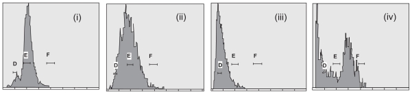
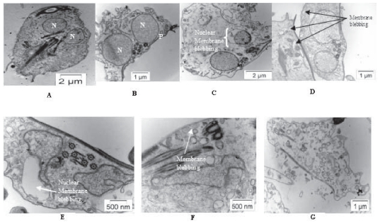
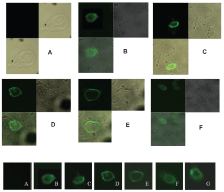
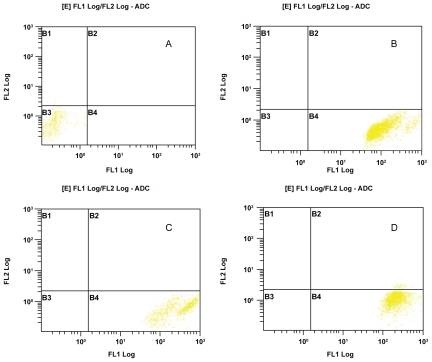
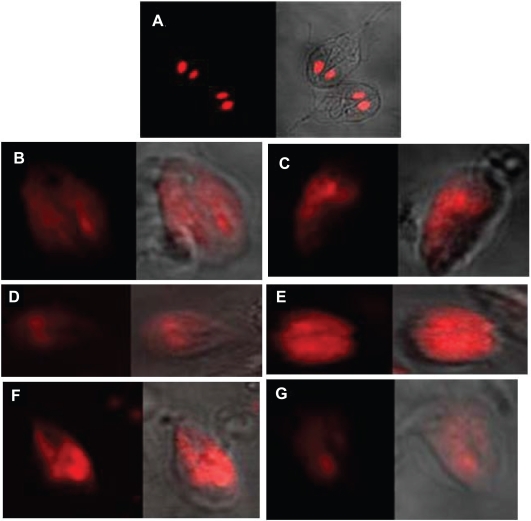
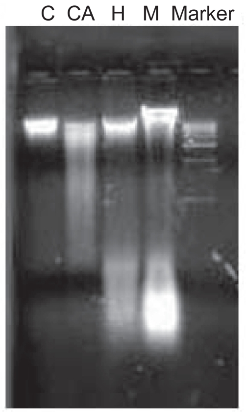
Giardia lamblia is a microaerophilic human gastrointestinal parasite and considered as an early-diverged eukaryote. In vitro oxidative stress generation plays a significant role in cell cycle progression and cell death of this parasite. In the present study hydrogen peroxide, metronidazole, and a modified growth medium without cysteine and ascorbic acid have been chosen as oxidative stress-inducing agents. Cell cycle progression has been found to be regulated by different types of oxidative stresses. Apoptosis is not an established pathway in Giardia, which is devoid of ideal mitochondria, but in the present investigation, apoptosis-like programmed cell death has been found by the experiments like AnnexinV-FITC assay, DNA fragmentation pattern, etc. On the contrary, Caspase-9 assay, which confirms the caspase-mediated apoptotic pathway, has been found to be negative in all the stress conditions. Protease inhibitor assay confirmed that, even in absence of any proteases, programmed cell death does occur in this primitive eukaryote. All these results signify a novel pathway of programmed suicidal death in Giardia lamblia under oxidative stress. This is the first demonstration of protease-independent programmed cell death regulation in Giardia exclusive for its own specialties.
Keywords: Giardia lamblia; apoptosis; early branching eukaryotes; oxidative stress; programmed cell death; reactive oxygen species.
Figures
Similar articles
-
Pyruvate Protects Giardia Trophozoites from Cysteine-Ascorbate Deprived Medium Induced Cytotoxicity.Korean J Parasitol. 2018 Feb;56(1):1-9. doi: 10.3347/kjp.2018.56.1.1. Epub 2018 Feb 28. Korean J Parasitol. 2018. PMID: 29529844 Free PMC article.
-
Differential gene expression in Giardia lamblia under oxidative stress: significance in eukaryotic evolution.Gene. 2014 Feb 10;535(2):131-9. doi: 10.1016/j.gene.2013.11.048. Epub 2013 Dec 7. Gene. 2014. PMID: 24321693
-
Programmed cell death in Giardia.Parasitology. 2012 Jun;139(7):894-903. doi: 10.1017/S003118201200011X. Epub 2012 Mar 12. Parasitology. 2012. PMID: 22405231
-
Anaerobic bacterial metabolism in the ancient eukaryote Giardia duodenalis.Int J Parasitol. 1998 Jan;28(1):149-64. doi: 10.1016/s0020-7519(97)00172-0. Int J Parasitol. 1998. PMID: 9504342 Review.
-
Molecular basis of defence against oxidative stress in Entamoeba histolytica and Giardia lamblia.Microbes Infect. 1999 Apr;1(5):385-94. doi: 10.1016/s1286-4579(99)80055-0. Microbes Infect. 1999. PMID: 10602671 Review.
Cited by
-
Apoptotic markers in protozoan parasites.Parasit Vectors. 2010 Nov 9;3:104. doi: 10.1186/1756-3305-3-104. Parasit Vectors. 2010. PMID: 21062457 Free PMC article.
-
Regulation of a Myb transcription factor by cyclin-dependent kinase 2 in Giardia lamblia.J Biol Chem. 2012 Feb 3;287(6):3733-50. doi: 10.1074/jbc.M111.298893. Epub 2011 Dec 13. J Biol Chem. 2012. PMID: 22167200 Free PMC article.
-
On the molecular and cellular effects of omeprazole to further support its effectiveness as an antigiardial drug.Sci Rep. 2019 Jun 20;9(1):8922. doi: 10.1038/s41598-019-45529-w. Sci Rep. 2019. PMID: 31222100 Free PMC article.
-
An antioxidant response is involved in resistance of Giardia duodenalis to albendazole.Front Microbiol. 2015 Apr 10;6:286. doi: 10.3389/fmicb.2015.00286. eCollection 2015. Front Microbiol. 2015. PMID: 25914688 Free PMC article.
-
Pyruvate Protects Giardia Trophozoites from Cysteine-Ascorbate Deprived Medium Induced Cytotoxicity.Korean J Parasitol. 2018 Feb;56(1):1-9. doi: 10.3347/kjp.2018.56.1.1. Epub 2018 Feb 28. Korean J Parasitol. 2018. PMID: 29529844 Free PMC article.
References
-
- Acker H. Mechanisms and meaning of cellular oxygen sensing in the organism. Respir Physiol. 1994;95(1):1–10. - PubMed
-
- Sen CK, Packer L. Antioxidant and redox regulation of gene transcription. FASEB J. 1996;10(7):709–720. - PubMed
-
- Atkinson HJ. Respiration in nematodes. In: Zuckerman BM, editor. Nematodes as Biological Models. New York, NY: Academic Press; 1980. pp. 116–142.
Publication types
LinkOut - more resources
Full Text Sources
