

Musculoskeletal complications of hemophilia
- PMID: 19921342
- PMCID: PMC2821487
- DOI: 10.1007/s11420-009-9140-9
Musculoskeletal complications of hemophilia
Abstract
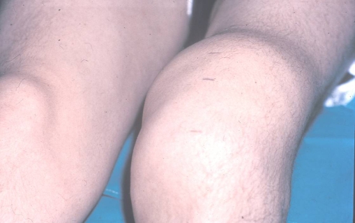
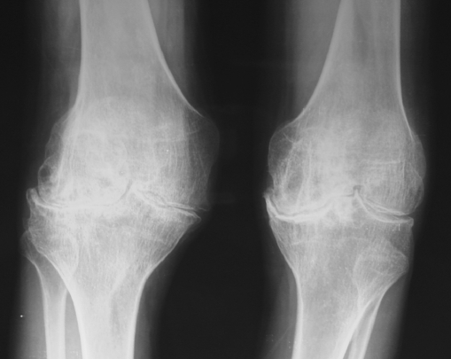
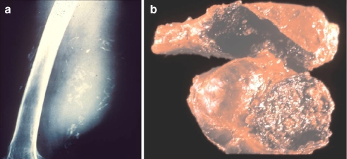
The most important clinical strategy for management of patients with hemophilia is the avoidance of recurrent hemarthroses by means of continuous, intravenous hematological prophylaxis. When only intravenous on-demand hematological treatment is available, frequent evaluations are necessary for the early diagnosis and treatment of episodes of intra-articular bleeding. The natural history of the disease in patients with poorly controlled intra-articular bleeding is the development of chronic synovitis and, later, multi-articular hemophilic arthropathy. Once arthropathy develops, the functional prognosis is poor. Treatment of these patients should be conducted through a comprehensive program by a multidisciplinary hemophilia unit. Although continuous prophylaxis can avoid the development of the orthopedic complications of hemophilia still seen in the twenty-first century, such a goal has not, so far, been achieved even in developed countries. Therefore, many different surgical procedures such as arthrocentesis, radiosynoviorthesis (radiosynovectomy) (yttrium-90, rhenium-186), tendon lengthenings, alignment osteotomies, joint arthroplasties, removal of pseudotumours, and fixation of fractures are still frequently needed in the care of these patients.
Figures
References
-
- Rodriguez-Merchan EC. Management of orthopedic complications of haemophilia. J. Bone Joint Surg. (Br) 1998;80B:191–196. - PubMed
LinkOut - more resources
Full Text Sources
Other Literature Sources
