Characterizing breast cancer xenograft epidermal growth factor receptor expression by using near-infrared optical imaging
- PMID: 19922304
- PMCID: PMC3618993
- DOI: 10.3109/02841850903008800
Characterizing breast cancer xenograft epidermal growth factor receptor expression by using near-infrared optical imaging
Abstract
Background: Epidermal growth factor receptor (EGFR) overexpression is associated with several key features of cancer development and growth. Therefore, EGFR is a very promising biological target for tumor diagnosis and anticancer therapy. Characterization of EGFR expression is important for clinicians to select patients for EGFR-targeted therapy and evaluate therapeutic effects.
Purpose: To investigate whether near-infrared (NIR) fluorescent dye Cy5.5-labeled anti-EGFR monoclonal antibody Erbitux can characterize EGFR expression level in MDA-MB-231 and MCF-7 breast cancer xenografts using an in vivo NIR imaging method.
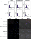
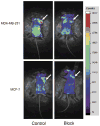
Material and methods: A fluorochrome probe was designed by coupling Cy5.5 to Erbitux through acidylation, and the fluorescence property of the Erbitux-Cy5.5 conjugate was characterized by fluorospectroscopy. Flow cytometry and laser confocal microscopy were used to test the EGFR specificity of the antibody probe in vitro. Erbitux-Cy5.5 was also injected intravenously into immune-deficient mice bearing MDA-MB-231 or MCF-7 tumors. Whole-body and region-of-interest fluorescence images were acquired and analyzed. The EGFR expression was also analyzed and confirmed by immunohistochemical assay.
Results: The maximum excitation/emission wavelength for the Erbitux-Cy5.5 probe was 674/697 nm, similar to that of free Cy5.5 (674/712 nm). Confocal microscopy confirmed receptor-specific uptake in MDA-MB-231 and MCF-7 cells. In flow cytometry probe specificity assay, Erbitux-Cy5.5 showed a 9.32-fold higher affinity for MDA-MB-231 than MCF-7 cells. In vivo NIR imaging also indicated specific uptake in EGFR-positive tumors. Probe uptake rate and maximum intake dose in MDA-MB-231 were significantly higher than those in MCF-7 xenografts (P < 0.001). Immunohistochemical staining confirmed the in vivo imaging results, showing differentiated EGFR expression in MDA-MB-231 (+ + +) and MCF-7 (+) tumor tissues.
Conclusion: Erbitux-Cy5.5 may be used as a specific NIR contrast agent for the noninvasive characterization of EGFR expression level in breast cancer xenografts.
Conflict of interest statement
Figures


Comment in
-
Molecular imaging: a potential new tool for early detection and monitoring of targeted breast cancer therapy.Acta Radiol. 2009 Dec;50(10):1092-3. doi: 10.3109/02841850903269188. Acta Radiol. 2009. PMID: 19922302 No abstract available.
Similar articles
-
Near-infrared optical imaging of epidermal growth factor receptor in breast cancer xenografts.Cancer Res. 2003 Nov 15;63(22):7870-5. Cancer Res. 2003. PMID: 14633715
-
Cy5.5-labeled Affibody molecule for near-infrared fluorescent optical imaging of epidermal growth factor receptor positive tumors.J Biomed Opt. 2010 May-Jun;15(3):036007. doi: 10.1117/1.3432738. J Biomed Opt. 2010. PMID: 20615009
-
99mTc-Hydrazinonicotinamide epidermal growth factor-polyethylene glycol-quantum dot imaging allows quantification of breast cancer epidermal growth factor receptor expression and monitors receptor downregulation in response to cetuximab therapy.J Nucl Med. 2011 Sep;52(9):1457-64. doi: 10.2967/jnumed.111.087619. Epub 2011 Aug 17. J Nucl Med. 2011. PMID: 21849406
-
IRDye 800-Anti–epidermal growth factor receptor Affibody.2010 Jun 17 [updated 2010 Sep 9]. In: Molecular Imaging and Contrast Agent Database (MICAD) [Internet]. Bethesda (MD): National Center for Biotechnology Information (US); 2004–2013. 2010 Jun 17 [updated 2010 Sep 9]. In: Molecular Imaging and Contrast Agent Database (MICAD) [Internet]. Bethesda (MD): National Center for Biotechnology Information (US); 2004–2013. PMID: 20845547 Free Books & Documents. Review.
-
QD800-Anti-epidermal growth factor receptor monoclonal antibody nanoparticles.2013 Jan 6 [updated 2013 Apr 4]. In: Molecular Imaging and Contrast Agent Database (MICAD) [Internet]. Bethesda (MD): National Center for Biotechnology Information (US); 2004–2013. 2013 Jan 6 [updated 2013 Apr 4]. In: Molecular Imaging and Contrast Agent Database (MICAD) [Internet]. Bethesda (MD): National Center for Biotechnology Information (US); 2004–2013. PMID: 23586108 Free Books & Documents. Review.
Cited by
-
Near infrared imaging of EGFR of oral squamous cell carcinoma in mice administered arsenic trioxide.PLoS One. 2012;7(9):e46255. doi: 10.1371/journal.pone.0046255. Epub 2012 Sep 28. PLoS One. 2012. PMID: 23029451 Free PMC article.
-
Detection and imaging of aggressive cancer cells using an epidermal growth factor receptor (EGFR)-targeted filamentous plant virus-based nanoparticle.Bioconjug Chem. 2015 Feb 18;26(2):262-269. doi: 10.1021/bc500545z. Epub 2015 Feb 5. Bioconjug Chem. 2015. PMID: 25611133 Free PMC article.
-
Inert coupling of IRDye800CW to monoclonal antibodies for clinical optical imaging of tumor targets.EJNMMI Res. 2011 Dec 1;1(1):31. doi: 10.1186/2191-219X-1-31. EJNMMI Res. 2011. PMID: 22214225 Free PMC article.
-
Recent trends in antibody-based oncologic imaging.Cancer Lett. 2012 Feb 28;315(2):97-111. doi: 10.1016/j.canlet.2011.10.017. Epub 2011 Oct 20. Cancer Lett. 2012. PMID: 22104729 Free PMC article. Review.
-
Near-infrared molecular probes for in vivo imaging.Curr Protoc Cytom. 2012 Apr;Chapter 12:Unit12.27. doi: 10.1002/0471142956.cy1227s60. Curr Protoc Cytom. 2012. PMID: 22470154 Free PMC article.
References
-
- Reilly RM, Kiarash R, Sandhu J, Lee YW, Cameron RG, Hendler A, et al. A comparison of EGF and MAb 528 labeled with 111In for imaging human breast cancer. J Nucl Med. 2000;41:903–11. - PubMed
-
- Klijn JG, Berns PM, Schmitz PI, Foekens JA. The clinical significance of epidermal growth factor receptor (EGF-R) in human breast cancer: a review on 5232 patients. Endocr Rev. 1992;13:3–17. - PubMed
-
- Ciardiello F, Tortora G. Anti-epidermal growth factor receptor drugs in cancer therapy. Expert Opin Investig Drugs. 2002;11:755–68. - PubMed
-
- Baselga J. Targeting tyrosine kinases in cancer: the second wave. Science. 2006;312:1175–8. - PubMed
-
- Blume-Jensen P, Hunter T. Oncogenic kinase signalling. Nature. 2001;411:355–65. - PubMed
Publication types
MeSH terms
Substances
Grants and funding
LinkOut - more resources
Full Text Sources
Medical
Research Materials
Miscellaneous
