

The ceramide-1-phosphate analogue PCERA-1 modulates tumour necrosis factor-alpha and interleukin-10 production in macrophages via the cAMP-PKA-CREB pathway in a GTP-dependent manner
- PMID: 19922425
- PMCID: PMC2826682
- DOI: 10.1111/j.1365-2567.2009.03188.x
The ceramide-1-phosphate analogue PCERA-1 modulates tumour necrosis factor-alpha and interleukin-10 production in macrophages via the cAMP-PKA-CREB pathway in a GTP-dependent manner
Abstract
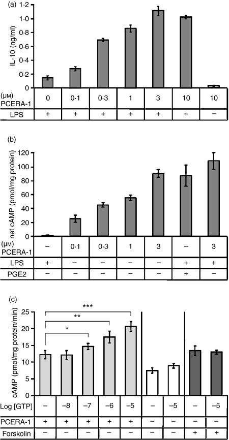
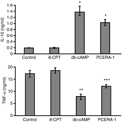
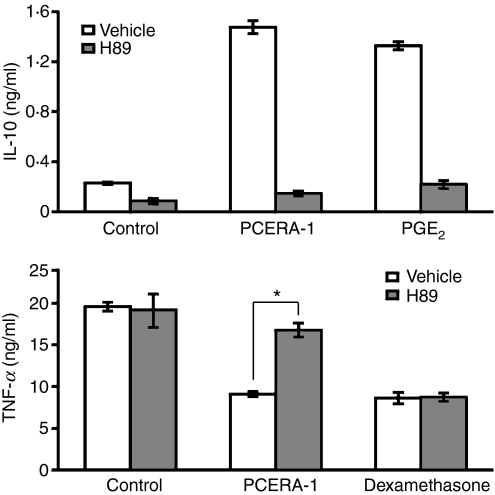
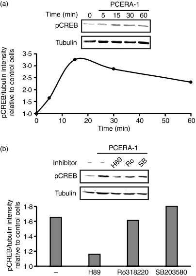
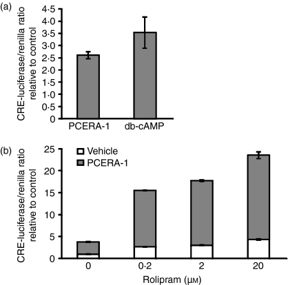
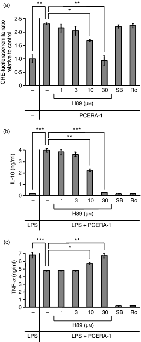
The synthetic phospho-ceramide analogue-1 (PCERA-1) down-regulates production of the pro-inflammatory cytokine tumour necrosis factor-alpha (TNF-alpha) and up-regulates production of the anti-inflammatory cytokine interleukin-10 (IL-10) in lipopolysaccharide (LPS) -stimulated macrophages. We have previously reported that PCERA-1 increases cyclic adenosine monophosphate (cAMP) levels. The objective of this study was to delineate the signalling pathway leading from PCERA-1 via cAMP to modulation of TNF-alpha and IL-10 production. We show here that PCERA-1 elevates intra-cellular cAMP level in a guanosine triphosphate-dependent manner in RAW264.7 macrophages. The cell-permeable dibutyryl cAMP was able to mimic the effects of PCERA-1 on cytokine production, whereas 8-chloro-phenylthio-methyladenosine-cAMP, which specifically activates the exchange protein directly activated by cAMP (EPAC) but not protein kinase A (PKA), failed to mimic PCERA-1 activities. Consistently, the PKA inhibitor H89 efficiently blocked PCERA-1-driven cytokine modulation as well as PCERA-1-stimulated phosphorylation of cAMP response element binding protein (CREB) on Ser-133. Finally, PCERA-1 activated cAMP-responsive transcription of a luciferase reporter, in synergism with the phosphodiesterase (PDE)-4 inhibitor rolipram. Our results suggest that PCERA-1 activates a G(s) protein-coupled receptor, leading to elevation of cAMP, which acts via the PKA-CREB pathway to promote TNF-alpha suppression and IL-10 induction in LPS-stimulated macrophages. Identification of the PCERA-1 receptor is expected to set up a new target for development of novel anti-inflammatory drugs.
Figures
Similar articles
-
Exogenous ceramide-1-phosphate (C1P) and phospho-ceramide analogue-1 (PCERA-1) regulate key macrophage activities via distinct receptors.Immunol Lett. 2016 Jan;169:73-81. doi: 10.1016/j.imlet.2015.12.001. Epub 2015 Dec 3. Immunol Lett. 2016. PMID: 26656944 Free PMC article.
-
Synergistic IL-10 induction by LPS and the ceramide-1-phosphate analog PCERA-1 is mediated by the cAMP and p38 MAP kinase pathways.Mol Immunol. 2009 Jun;46(10):1979-87. doi: 10.1016/j.molimm.2009.03.009. Epub 2009 Apr 10. Mol Immunol. 2009. PMID: 19362373
-
A ceramide-1-phosphate analogue, PCERA-1, simultaneously suppresses tumour necrosis factor-alpha and induces interleukin-10 production in activated macrophages.Immunology. 2009 May;127(1):103-15. doi: 10.1111/j.1365-2567.2008.02928.x. Immunology. 2009. PMID: 18793216 Free PMC article.
-
Cyclic nucleotide signaling changes associated with normal aging and age-related diseases of the brain.Cell Signal. 2018 Jan;42:281-291. doi: 10.1016/j.cellsig.2017.11.004. Epub 2017 Nov 23. Cell Signal. 2018. PMID: 29175000 Free PMC article. Review.
-
From membrane to nucleus: A three-wave hypothesis of cAMP signaling.J Biol Chem. 2024 Jan;300(1):105497. doi: 10.1016/j.jbc.2023.105497. Epub 2023 Nov 26. J Biol Chem. 2024. PMID: 38016514 Free PMC article. Review.
Cited by
-
Neosaxitoxin Inhibits the Expression of Inflammation Markers of the M1 Phenotype in Macrophages.Mar Drugs. 2020 May 27;18(6):283. doi: 10.3390/md18060283. Mar Drugs. 2020. PMID: 32471037 Free PMC article.
-
PGE(2) induces macrophage IL-10 production and a regulatory-like phenotype via a protein kinase A-SIK-CRTC3 pathway.J Immunol. 2013 Jan 15;190(2):565-77. doi: 10.4049/jimmunol.1202462. Epub 2012 Dec 14. J Immunol. 2013. PMID: 23241891 Free PMC article.
-
The expression of thioredoxin-1 in acute epinephrine stressed mice.Cell Stress Chaperones. 2016 Sep;21(5):935-41. doi: 10.1007/s12192-016-0722-4. Epub 2016 Aug 11. Cell Stress Chaperones. 2016. PMID: 27511023 Free PMC article.
-
Exogenous ceramide-1-phosphate (C1P) and phospho-ceramide analogue-1 (PCERA-1) regulate key macrophage activities via distinct receptors.Immunol Lett. 2016 Jan;169:73-81. doi: 10.1016/j.imlet.2015.12.001. Epub 2015 Dec 3. Immunol Lett. 2016. PMID: 26656944 Free PMC article.
-
The cyclic AMP signaling pathway: Exploring targets for successful drug discovery (Review).Mol Med Rep. 2016 May;13(5):3715-23. doi: 10.3892/mmr.2016.5005. Epub 2016 Mar 18. Mol Med Rep. 2016. PMID: 27035868 Free PMC article. Review.
References
-
- Beutler B. Innate immunity: an overview. Mol Immunol. 2004;40:845–59. - PubMed
-
- Szelenyi J, Kiss JP, Puskas E, Szelenyi M, Vizi ES. Contribution of differently localized alpha 2- and beta-adrenoceptors in the modulation of TNF-alpha and IL-10 production in endotoxemic mice. Ann N Y Acad Sci. 2000;917:145–53. - PubMed
-
- Takano M, Nishimura H, Kimura Y, Washizu J, Mokuno Y, Nimura Y, Yoshikai Y. Prostaglandin E2 protects against liver injury after Escherichia coli infection but hampers the resolution of the infection in mice. J Immunol. 1998;161:3019–25. - PubMed
-
- Kast RE. Tumor necrosis factor has positive and negative self regulatory feed back cycles centered around cAMP. Int J Immunopharmacol. 2000;22:1001–6. - PubMed
-
- Zidek Z. Adenosine – cyclic AMP pathways and cytokine expression. Eur Cytokine Netw. 1999;10:319–28. - PubMed
Publication types
MeSH terms
Substances
LinkOut - more resources
Full Text Sources
