. 2009 Nov 17;2(1):56.
doi: 10.1186/1756-3305-2-56.

Distribution of Brugia malayi larvae and DNA in vector and non-vector mosquitoes: implications for molecular diagnostics

Affiliations

Distribution of Brugia malayi larvae and DNA in vector and non-vector mosquitoes: implications for molecular diagnostics

Sara M Erickson et al. Parasit Vectors. .

Abstract

Background: The purpose of this study was to extend prior studies of molecular detection of Brugia malayi DNA in vector (Aedes aegypti- Liverpool) and non-vector (Culex pipiens) mosquitoes at different times after ingestion of infected blood.

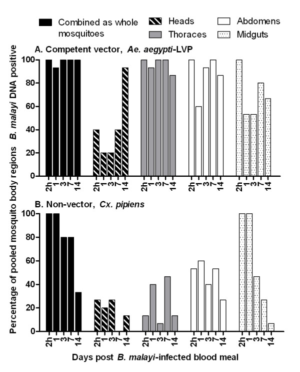
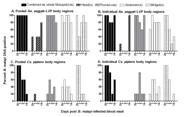
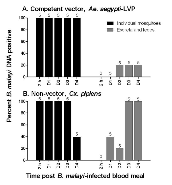
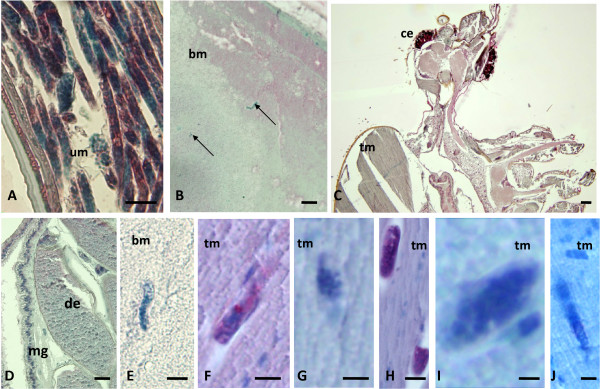
Results: Parasite DNA was detected over a two week time course in 96% of pooled thoraces of vector mosquitoes. In contrast, parasite DNA was detected in only 24% of thorax pools from non-vectors; parasite DNA was detected in 56% of midgut pools and 47% of abdomen pools from non-vectors. Parasite DNA was detected in vectors in the head immediately after the blood meal and after 14 days. Parasite DNA was also detected in feces and excreta of the vector and non-vector mosquitoes which could potentially confound results obtained with field samples. However, co-housing experiments failed to demonstrate transfer of parasite DNA from infected to non-infected mosquitoes. Parasites were also visualized in mosquito tissues by immunohistololgy using an antibody to the recombinant filarial antigen Bm14. Parasite larvae were detected consistently after mf ingestion in Ae. aegypti- Liverpool. Infectious L3s were seen in the head, thorax and abdomen of vector mosquitoes 14 days after Mf ingestion. In contrast, parasites were only detected by histology shortly after the blood meal in Cx. pipiens, and these were not labeled by the antibody.

Conclusion: This study provides new information on the distribution of filarial parasites and parasite DNA in vector and non-vector mosquitoes. This information should be useful for those involved in designing and interpreting molecular xenomonitoring studies.


Figures

Figure 1
Figure 1
Prevalence of B. malayi DNA in pooled samples of experimentally infected mosquitoes by body part (head, thorax, abdomen, and midgut) at different times post ingestion of microfilaremic blood. A Ae. aegypti-LVP, competent B. malayi vector B Cx. pipiens, B. malayi non-vector.
Figure 2
Figure 2
Comparison of B. malayi DNA prevalence in pooled and individually tested mosquito body parts. A Ae. aegypti-LVP body parts were tested in pools of four. B Ae. aegypti-LVP body parts individually tested for parasite DNA. C Cx. pipiens body parts tested in pools of four. D Cx. pipiens body parts individually tested for B. malayi DNA.
Figure 3
Figure 3
Detection of B. malayi DNA in mosquito feces. A Prevalence of parasite DNA in individually housed Ae. aegypti-LVP and their feces. B Prevalence of parasite DNA in individually housed Cx. pipiens and their feces. Sample number is indicated above each bar.
Figure 4
Figure 4
Immunohistological detection of B. malayi larvae in Ae. aegypti-LVP using polyclonal antibody to recombinant antigen Bm14. A Non-stained mf (arrow) in the midgut at 1 DPI: B Longitudinal section of a stained L1 in the thorax muscles at 1 DPI: C Cross-section of a stained larva at 3 DPI: D Multiple cross-sections of strongly labeled L2s (arrow) at 7 DPI in the thorax. E Cross-sections of two strongly labeled L2s with body cavity and developing intestine at 7 DPI: F Overview of the head with 4 cross-sections of strongly labeled L3s (arrow) at 14 DPI: G Cross-section of 2 well stained larvae close to the mosquito eye. H Labeled L3 at 14 DPI in the mosquito mouthparts. I Multiple strongly labeled L3s (arrows) in the thorax. J Strongly labeled L3 in the abdomen. (bm, blood meal; ce, compound eye; de, developing egg; tm, thorax muscles; mg, midgut; um, uterus membrane). Scale bars: A-C, E 25 μm; D, F-J 50 μm.
Figure 5
Figure 5
Immunohistological detection of B. malayi larvae in the uterus of adult B. malayi or in experimentally infected mosquitoes using polyclonal antibody to recombinant antigen Bm14. A The antibody labeled intra-uterine, stretched mf in an adult female B. malayi worm. B Multiple unlabeled mf in the midgut of Cx. pipiens at 2 h PI: C Overview of the head and parts of the thorax at 14 DPI negative for B. malayi larvae. D Abdomen of Cx. pipiens at 3 DPI without any visible developing B. malayi larvae (labeled or unlabeled). E Magnified, unlabeled mf in the midgut of Cx. pipiens at 2 h PI: F Section of a labeled L1 in the thorax muscles of Ae. aegypti-LVP at 1 DPI: G Section of an unlabeled L1 in the thorax of Ae. aegypti-RKF at 1 DPI: H Two sections of labeled larvae in the thorax of Ae. aegypti-LVP at 3 DPI: I Section of an unlabeled larva in Ae. aegypti-RKF at 3 DPI. J Section of an unlabeled larva in Ae. aegypti-RKF at 7 DPI (compare Fig. 4D). Scale bars: A-D 25 μm; E-F 10 μm.

References

    1. WHO. Global programme to eliminate lymphatic filariasis. Wkly Epidemiol Rec. 2007;82:361–380. - PubMed
    1. Michael E, Malecela MN, Zervos M, Kazura JW. Global eradication of lymphatic filariasis: the value of chronic disease control in parasite elimination programmes. PLoS ONE. 2008;3:e2936. doi: 10.1371/journal.pone.0002936. - DOI - PMC - PubMed
    1. Kyelem D, Biswas G, Bockarie MJ, Bradley MH, El-Setouhy M, Fischer PU, Henderson RH, Kazura JW, Lammie PJ, Njenga SM, Ottesen EA, Ramaiah KD, Richards FO, Weil GJ, Williams SA. Determinants of success in national programs to eliminate lymphatic filariasis: a perspective identifying essential elements and research needs. Am J Trop Med Hyg. 2008;79:480–484. - PMC - PubMed
    1. Ramzy RM, El Setouhy M, Helmy H, Ahmed ES, Abd Elaziz KM, Farid HA, Shannon WD, Weil GJ. Effect of yearly mass drug administration with diethylcarbamazine and albendazole on bancroftian filariasis in Egypt: a comprehensive assessment. Lancet. 2006;367:992–999. doi: 10.1016/S0140-6736(06)68426-2. - DOI - PubMed
    1. Weil GJ, Ramzy RM. Diagnostic tools for filariasis elimination programs. Trends Parasitol. 2007;23:78–82. doi: 10.1016/j.pt.2006.12.001. - DOI - PubMed

LinkOut - more resources