Comparative Study
. 2009 Nov 17:9:18.
doi: 10.1186/1472-6904-9-18.

In vivo administration of BL-3050: highly stable engineered PON1-HDL complexes

Affiliations
Comparative Study

In vivo administration of BL-3050: highly stable engineered PON1-HDL complexes

Leonid Gaidukov et al. BMC Clin Pharmacol. .

Abstract

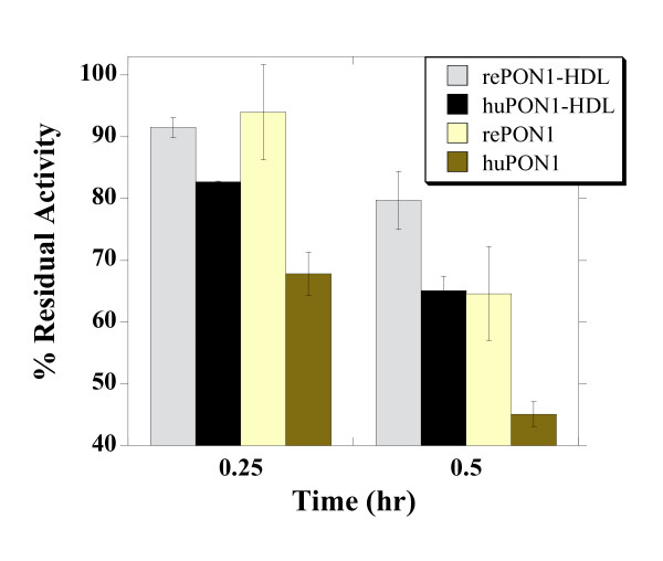
Background: Serum paraoxonase (PON1) is a high density lipoprotein (HDL)-associated enzyme involved in organophosphate (OP) degradation and prevention of atherosclerosis. PON1 comprises a potential candidate for in vivo therapeutics, as an anti-atherogenic agent, and for detoxification of pesticides and nerve agents. Because human PON1 exhibits limited stability, engineered, recombinant PON1 (rePON1) variants that were designed for higher reactivity, solubility, stability, and bacterial expression, are candidates for treatment. This work addresses the feasibility of in vivo administration of rePON1, and its HDL complex, as a potentially therapeutic agent dubbed BL-3050.

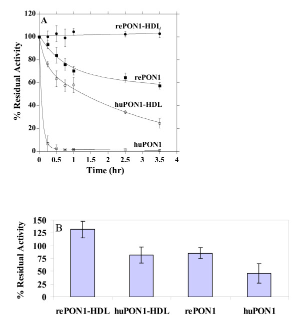
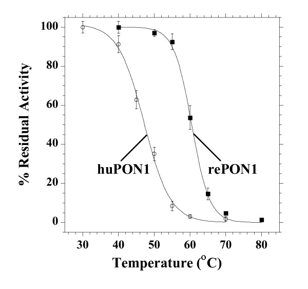
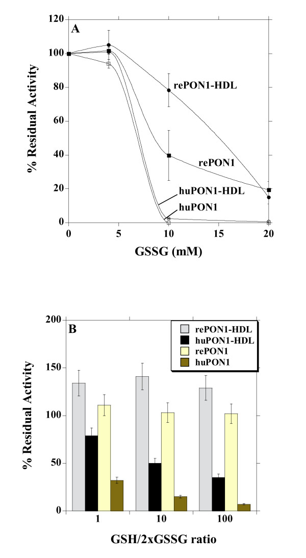
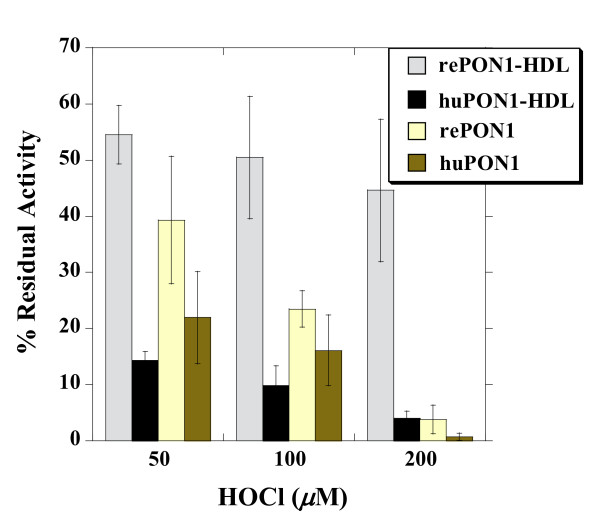
Methods: For stability studies we applied different challenges related to the in vivo disfunctionalization of HDL and PON1 and tested for inactivation of PON1's activity. We applied acute, repetitive administrations of BL-3050 in mice to assess its toxicity and adverse immune responses. The in vivo efficacy of recombinant PON1 and BL-3050 were tested with an animal model of chlorpyrifos-oxon poisoning.

Results: Inactivation studies show significantly improved in vitro lifespan of the engineered rePON1 relative to human PON1. Significant sequence changes relative to human PON1 might hamper the in vivo applicability of BL-3050 due to adverse immune responses. However, we observed no toxic effects in mice subjected to repetitive administration of BL-3050, suggesting that BL-3050 could be safely used. To further evaluate the activity of BL-3050 in vivo, we applied an animal model that mimics human organophosphate poisoning. In these studies, a significant advantages of rePON1 and BL-3050 (>87.5% survival versus <37.5% in the control groups) was observed. Furthermore, BL-3050 and rePON1 were superior to the conventional treatment of atropine-2-PAM as a prophylactic treatment for OP poisoning.

Conclusion: In vitro and in vivo data described here demonstrate the potential advantages of rePON1 and BL-3050 for treatment of OP toxicity and chronic cardiovascular diseases like atherosclerosis. The in vivo data also suggest that rePON1 and BL-3050 are stable and safe, and could be used for acute, and possibly repeated treatments, with no adverse effects.


Figures

Figure 1
Figure 1
Inactivation of the recombinant and human PON1, and PON1-HDL complexes in the presence of calcium chelator (A) or in buffer (B). Data represents mean ± SD of at least 3 independent experiments. A. Inactivation kinetics of rePON1, rePON1-HDL, huPON1, and huPON1-HDL (huHDL) by EDTA (4 mM) and β-mercaptoethanol (8 mM) at 37°C. Residual activity at various time points was determined by initial rates of phenyl acetate hydrolysis and plotted as percent of the activity at time zero. Data were fitted to a single exponential for rePON1-HDL, and to double-exponentials for the remaining samples [35]. B. Inactivation of rePON1, rePON1-HDL, huPON1, and huPON1-HDL (huHDL), in PBS buffer with 1 mM CaCl2. Residual PON1 activity after 24 hrs incubation at 37°C is presented as percentage of the initial activity.
Figure 2
Figure 2
Heat inactivation of rePON1 and huPON1. Purified enzymes diluted in TBS with 1 mM CaCl2 were incubated at the range of temperatures (25 - 80°C) for 30 mins, and residual PON1 activity was determined. Data were fitted to the sigmoidal decay function to give Tm values of 47.2°C for huPON1 and 60.4°C for rePON1 (the corresponding m values are 0.273 and 0.399; see Methods). Each point represents mean ± SD of 3 experiments.
Figure 3
Figure 3
Inactivation of the recombinant and human PON1, and PON1-HDL complexes by glutathione. Data represents mean ± SD of at least 3 independent experiments. A. Recombinant and human PON1, and the corresponding HDL complexes, were incubated with increasing concentrations of GSSG at 37°C. Residual PON1 activity was determined after 40 min of incubation, and plotted as percentage of the initial activity. B. Recombinant and human PON1, and the corresponding HDL complexes, were incubated with various GSH/GSSG ratios (the ratios are expressed as the ratio of glutathione equivalents, i.e., the ratio of GSH to 2xGSSG) whilst keeping the total glutathione concentration at 10 mM. Residual PON1 activity was determined after 40 min of incubation, and plotted as percentage of the initial activity.
Figure 4
Figure 4
Inactivation of the recombinant and human PON1, and PON1-HDL complexes by hypochlorite. Recombinant and human PON1, and the corresponding HDL complexes, were incubated with increasing concentrations of hypochlorite at 37°C. Residual PON1 activity was determined after 24 hrs incubation, and plotted as percentage of the initial activity. Residual PON1 activity in the absence of hypochlorite remained largely unchanged for all the samples (≥ 75%). Data represents mean ± SD of 4 independent experiments.
Figure 5
Figure 5
Inactivation of recombinant and human PON1, and PON1-HDL complexes by tetranitromethane. PON1 samples were incubated with 1 mM of TNM at 37°C. Residual PON1 activity was determined after 0.25 and 0.5 hrs of incubation, and plotted as percentage of the initial activity. Residual PON1 activity with no addition of TNM remained unchanged for all the samples (~100%). Data represents mean ± SD of 3 independent experiments performed in duplicates.

References

    1. Khersonsky O, Tawfik DS. Structure-Reactivity Studies of Serum Paraoxonase PON1 Suggest that Its Native Activity Is Lactonase. Biochemistry. 2005;44(16):6371–6382. doi: 10.1021/bi047440d. - DOI - PubMed
    1. Draganov DI, Teiber JF, Speelman A, Osawa Y, Sunahara R, La Du BN. Human paraoxonases (PON1, PON2, and PON3) are lactonases with overlapping and distinct substrate specificities. J Lipid Res. 2005;46(6):1239–1247. doi: 10.1194/jlr.M400511-JLR200. - DOI - PubMed
    1. Camps J, Marsillach J, Joven J. The paraoxonases: role in human diseases and methodological difficulties in measurement. Critical reviews in clinical laboratory sciences. 2009;46(2):83–106. doi: 10.1080/10408360802610878. - DOI - PubMed
    1. Sorenson RC, Bisgaier CL, Aviram M, Hsu C, Billecke S, La Du BN. Human serum Paraoxonase/Arylesterase's retained hydrophobic N-terminal leader sequence associates with HDLs by binding phospholipids: apolipoprotein A-I stabilizes activity. Arterioscler Thromb Vasc Biol. 1999;19(9):2214–2225. - PubMed
    1. Deakin S, Leviev I, Gomaraschi M, Calabresi L, Franceschini G, James RW. Enzymatically active paraoxonase-1 is located at the external membrane of producing cells and released by a high affinity, saturable, desorption mechanism. J Biol Chem. 2002;277(6):4301–4308. doi: 10.1074/jbc.M107440200. - DOI - PubMed

Publication types

MeSH terms

LinkOut - more resources