. 2009 Nov 18;10(1):112.
doi: 10.1186/1465-9921-10-112.

Impaired immune responses in the lungs of aged mice following influenza infection

Affiliations

Impaired immune responses in the lungs of aged mice following influenza infection

Franklin R Toapanta et al. Respir Res. .

Abstract

Background: Each year, influenza virus infection causes severe morbidity and mortality, particularly in the most susceptible groups including children, the elderly (>65 years-old) and people with chronic respiratory diseases. Among the several factors that contribute to the increased susceptibility in elderly populations are the higher prevalence of chronic diseases (e.g. diabetes) and the senescence of the immune system.

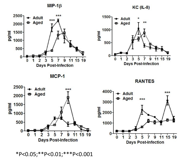
Methods: In this study, aged and adult mice were infected with sublethal doses of influenza virus (A/Puerto Rico/8/1934). Differences in weight loss, morbidity, virus titer and the kinetics of lung infiltration with cells of the innate and adaptive immune responses were analyzed. Additionally, the main cytokines and chemokines produced by these cells were also assayed.

Results: Compared to adult mice, aged mice had higher morbidity, lost weight more rapidly, and recovered more slowly from infection. There was a delay in the accumulation of granulocytic cells and conventional dendritic cells (cDCs), but not macrophages in the lungs of aged mice compared to adult animals. The delayed infiltration kinetics of APCs in aged animals correlated with alteration in their activation (CD40 expression), which also correlated with a delayed detection of cytokines and chemokines in lung homogenates. This was associated with retarded lung infiltration by natural killer (NK), CD4+ and CD8+ T-cells. Furthermore, the percentage of activated (CD69+) influenza-specific and IL-2 producer CD8+ T-cells was higher in adult mice compared to aged ones. Additionally, activation (CD69+) of adult B-cells was earlier and correlated with a quicker development of neutralizing antibodies in adult animals.

Conclusion: Overall, alterations in APC priming and activation lead to delayed production of cytokines and chemokines in the lungs that ultimately affected the infiltration of immune cells following influenza infection. This resulted in delayed activation of the adaptive immune response and subsequent delay in clearance of virus and prolonged illness in aged animals. Since the elderly are the fastest growing segment of the population in developed countries, a better understanding of the changes that occur in the immune system during the aging process is a priority for the development of new vaccines and adjuvants to improve the immune responses in this population.


Figures

Figure 1
Figure 1
Mouse Morbidity and Lung Virus Titer. Following infection with a sublethal dose of PR8 virus, mice were evaluated daily for weight loss (A) and increase in sickness score (B). Adult mice initial weight was significantly lower than aged animals (21.27 ± 0.2785 vs 24.93 ± 0.5157; P < 0.01). Aged animals (white squares) lost weight quicker, had a higher sickness score and recovered slower than adult mice (black circles). Mock infected animals (white circles -- adults; black squares - aged) did not lost weight or showed signs of disease. Influenza virus titer was evaluated in lung supernatants of adult (black bars) and aged (white bars) mice by plaquing on MDCK cells (C). Aged and adult mice had high virus titer in the lungs. Virus was recovered for a longer period of time in aged animals (C), which correlated with the delayed weight recovery (A) from aged animals and delayed reduction in sickness score (B). This suggested alterations in the immune system of elderly animals. Panels A and B are composites of three different experiments and panel C of 2 experiments. In each graph, the arithmetic mean ± SEM are displayed. The number of animals (N) used to generate each graph is displayed at the bottom of each panel. Stars indicate statistical difference between aged and naïve animals. *P < 0.05. **P < 0.01. ***P < 0.001.
Figure 2
Figure 2
Lung Cell Infiltration. Isolated lung cells were evaluated for viability by trypan blue exclusion (A). The number of cells was adjusted for the weight of the lungs and reported as cells per gram of tissue. Aged mice (white squares) showed a delay in the kinetics of lung cell. The graph shows cumulative data of three sets of experiments, a total of 9 mice were assayed at each time point, except for days 15 and 19, which contain 8 animals each. (B) Gating strategy used to analyze innate cells in the lungs. Initially cells were gated in the non-lymphocyte area (R1), this was followed by selection in the live gate (R2). Two APC populations (R3 and R4 gates) were selected based on CD11b and CD11c expression. R3 gate corresponds to cDCs (CD11chigh/CD11bhigh). Cells in R4 gate were further divided based on the diffential expression of Gr-1 (neutrophils (CD11bhigh/CD11clow/Gr1high) and lung macrophages (CD11bhigh/CD11clow/Gr1med). MHC class II and CD40 expression was analyzed in R3 and R6 gates. An average of 500,000 events was collected for the analysis of these populations. Stars indicate statistical difference between aged and naïve animals.*P < 0.05. **P < 0.01. ***P < 0.001.
Figure 3
Figure 3
Innate Cells in the Lungs. Naïve mice (day 0) had no statistically significant differences in the innate populations analyzed by flow cytometry (A); however, aged animals had the tendency to have a higher percentage of granulocytes. There was a delay in the kinetics of granulocyte and cDCs infiltration in the lungs of aged mice (white squares) (B and D), which suggested alterations in chemokine production. Lung macrophages did not show altered infiltration kinetics, but cleared more quickly from the lungs of adult mice (black circles) (C), which correlated with a quicker recovery from adult mice. Stars indicate statistical difference between aged and naïve animals.*P < 0.05. **P < 0.01. ***P < 0.001.
Figure 4
Figure 4
Innate Cells Activation. The expression of CD40 and MHC class II was assayed in lung macrophages and cDCs (R6 and R3 in Fig 2B, respectively). Example of expression of CD40 by these cells is shown in adult (first column) and aged (second column) animals at different time points (A and D). An overlay of the histograms is shown to compare differences in up-regulation (third column) of the marker. Panels B and C show upergulation of CD40 and MHC class II respectively. Panels B and C display the average of two different experiments, on which of 3 mice were assayed at each time point (a total of 6 animals per time point), bars represent SEM. Aged animals are represented in white squares and adult animals in black circles. Expression of CD40 on lung macrophages and cDCs was similar in adult and aged animals at day 0. Up-regulation of this marker was slower in aged animals and statistically significant differences were detected as early as day 3 (B and E). On the other hand, MHC class II expression at day 0 and up-regulation was similar between adult and aged animals in both lung macrophages and cDCs (C and F). For expression of activation markers an average of 500,000 events were collected. *P < 0.05. **P < 0.01. ***P < 0.001.
Figure 5
Figure 5
Pro-inflamatory and lymphocytic cytokines. The concentration of several pro-inflamatory and lymphocytic cytokines was determined in the supernatants of lung homogenates. IL-12p70 (NK and CD4 T-cell stimulant) was produced earlier and at significantly higher levels in adult mice (black circles) (A), which correlated with an earlier detection of IFN-γ in adult animals. IL-1β concentrations peaked at day 5 post-infection in adult mice (black circle), but steadily increased in aged mice (B). TNF-α and IL-1α concentrations increased similarly in both adult and aged mice until day 5 and then declined over the 19 days of observation in adult animals (black circles)(C and D). In contrast, these two cytokines continued to increase in aged mice (white squares) with significantly higher concentrations at day 7-9 post-infection (C and D). The lymphocytic cytokines, IFN-γ and IL-6, spiked earlier in adult mice (black circles) (E and F). The graphs represent cumulative results of two different experiments. The arithmetic mean (± SEM) of the cytokine(s) concentration of 4-6 mice assayed at each time point was plotted. Stars indicate statistical difference between aged and naïve animals.*P < 0.05. **P < 0.01. ***P < 0.001.
Figure 6
Figure 6
Chemokines. Several chemokines were determined in the lung supernatants of aged and adult mice infected with influenza virus. MIP-1β increased and peaked earlier in adult animals (black squares) (A). KC increased similarly in the lungs of adult (black circles) and aged (white squares) mice, however peaked earlier in adult animals (B). MCP-1 had a mild early peak in adult mice (black circles), while a higher delayed peak in aged animals (white squares) (C). There was a biphasic expression pattern of RANTES in adult mice (black circles) with a peak in concentrations at day 5 and 15 (D), while aged animals (white squares) had a very mild increase in this chemokine. The overall delayed production of chemokines correlated with delays in infiltration kinetics of innate and adaptive cells the lungs. The graphs represent cumulative results of two different experiments. The arithmetic mean (± SEM) concentration of 4-6 mice assayed at each time point was plotted. Stars indicate statistical difference between aged and naïve animals. *P < 0.05. **P < 0.01. ***P < 0.001.
Figure 7
Figure 7
Lymphocytic cells. Several innate and adaptive lymphocytic cells were analyzed in the lungs of aged and adult mice. CD4+ and CD8+ T-cells were more abundant in adult mice (black bars) at day 0 (A). Adult mice (black circles) had a significantly higher infiltration of NK (DX5) cells in the lungs (B). The kinetics of B-cell lung infiltration, on adult (black circles) and aged (white squares) mice were similar (C). Expression of the early activation marker CD69 by NK and B-cells was detected earlier and at a higher percentage in adult mice in (black circles) (E and F), which suggested that despite similar infiltration kinetics, adult B-cells started to produce antibodies earlier. This was confirmed later when HAI were performed (Fig. 8). The graphs represent cumulative results of two different experiments. The arithmetic means (± SEM) of the number of cells (C and D) or percentages (A, D and E) at each time point (4-6 mice per time point) were plotted. Stars indicate statistical difference between aged and naïve animals. *P < 0.05. **P < 0.01. ***P < 0.001.
Figure 8
Figure 8
Anti-Influenza Neutralizing Antibodies. Anti-influenza neutralizing antibodies titers were assayed in sera samples by HAI assay. Aged animals (black circles) developed antibodies at neutralizing titer (1:40 -- dotted line) quicker than aged mice (white squares). The arithmetic mean (± SEM) of the sera HAI titer at each time point was plotted. Six mice per time point were assayed, except at days 7 and 15, where 10 mice were assayed. Stars indicate statistical difference between aged and naïve animals. *P < 0.05. **P < 0.01. ***P < 0.001.
Figure 9
Figure 9
T-lymphocytes. Correlating with a delayed detection of cytokines and chemokines in the supernatants of lung homogenates (Figs. 5 and 6), CD4+ and CD8+ T-cell infiltration was delayed in the lungs of aged animals (white squares) (A and B). However, up-regulation of CD69 was similar in aged and adult animals (C and D), which suggested non-specific up-regulation of this marker by these cells. Confirming this, activated influenza specific cells were detected only after day 9 post-infection (E and F). Plots A to D represent cumulative results of two different experiments. The arithmetic means (± SEM) of the number of cells (A and B) or percentages (C and D) at each time point (4-6 mice per time point) were plotted. In panels E and F, 5 mice per time point were assayed bars represent the arithmetic mean (± SEM). Stars indicate statistical difference between aged and adult animals. *P < 0.05. **P < 0.01. ***P < 0.001.
Figure 10
Figure 10
Intracellular Cytokine Staining. IL-2 (A and B), TNF-α (C and D) and IFN-γ (E and F) production were assayed in CD8+ T-cells following stimulation with immunodominant influenza peptides at day 15. Even though not statistically significant differences between adult and aged animals were detected, adult animals had the tendency to have a higher percentage of cells producing these cytokines regardless of HA or NP stimualtion. Five to eight mice per time point were assayed. Bars represent the arithmetic mean (± SEM). Stars indicate statistical difference between aged and naïve animals. *P < 0.05. **P < 0.01. ***P < 0.001.

References

    1. Russell CA, Jones TC, Barr IG, Cox NJ, Garten RJ, Gregory V. et al.The global circulation of seasonal influenza A (H3N2) viruses. Science. 2008;320(5874):340–6. doi: 10.1126/science.1154137. - DOI - PubMed
    1. Stohr K. Influenza-WHO cares. Lancet Infect Dis. 2002;2(9):517. doi: 10.1016/S1473-3099(02)00366-3. - DOI - PubMed
    1. Yoshikawa TT. Important infections in elderly persons. West J Med. 1981;135(6):441–5. - PMC - PubMed
    1. Bender BS, Johnson MP, Small PA. Influenza in senescent mice: impaired cytotoxic T-lymphocyte activity is correlated with prolonged infection. Immunology. 1991;72(4):514–9. - PMC - PubMed
    1. Bender BS, Taylor SF, Zander DS, Cottey R. Pulmonary immune response of young and aged mice after influenza challenge. J Lab Clin Med. 1995;126(2):169–77. - PubMed

Publication types

MeSH terms