

Review
doi: 10.1016/j.semcdb.2009.11.011.
Epub 2009 Nov 13.
Multiscale simulation of protein mediated membrane remodeling
Affiliations
- PMID: 19922811
- PMCID: PMC2855739
- DOI: 10.1016/j.semcdb.2009.11.011
Item in Clipboard
Review
Multiscale simulation of protein mediated membrane remodeling
Semin Cell Dev Biol.
2010 Jun.
Abstract
Proteins interacting with membranes can result in substantial membrane deformations and curvatures. This effect is known in its broadest terms as membrane remodeling. This review article will survey current multiscale simulation methodologies that have been employed to examine protein mediated membrane remodeling.
(c) 2009 Elsevier Ltd. All rights reserved.
Figures
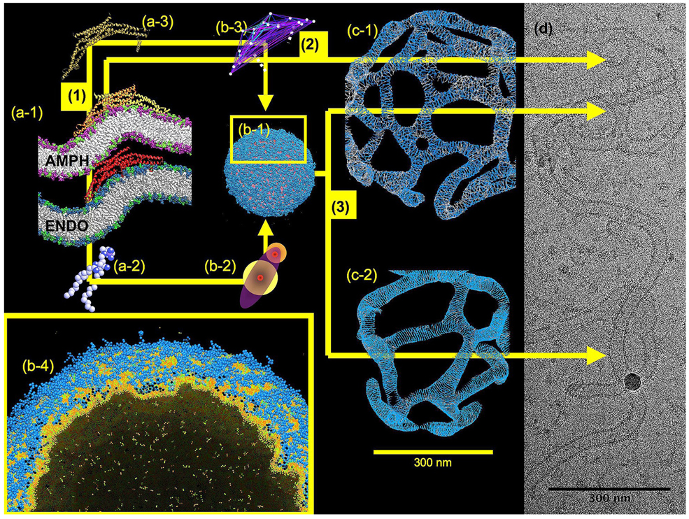
A schematic of the multiscale characteristics of protein mediated membrane remodeling. Images (a-1) to (a-3), (b-1) to (b-4), and then (c-1) and (c-2) give simulation snapshots of N-BAR domain driven membrane remodeling at the atomic, CG, and mesoscopic scales, respectively. The colored arrows designate different multiscale paths where by the simulations can ultimately connect with EM imaging (EM image (d) of amphiphysin tubulation courtesy of V. Unger). Path (1) connects image (a-1), the atomic level, with (b-1) the CG scale. In this path, lipids (a-2) and proteins (a-3) are systematically coarse-grained into much simpler objects (b-2) and (b-3), but still retain residual atomic-level information. An entire 200 nm diameter liposome composed of around half a million CG lipids undergoing the early stages of remodeling is shown in image (b-1). Path (2) directly connects the atomic with the mesoscopic scale, and is a methodology sometimes employed. This path requires fairly extensive phenomenological information. However, as path (2) connects to image (c-1) and (c-2), the mesoscopic representation, liposome remodeling of 500 nm diameter liposomes can be described over effectively very long (macroscopic) timescales. It is at this end point that direct comparisons with EM imaging can be achieved (image d). The two different mesoscopic images, (c-1) and (c-2), give an indication of the polymorphism of structures that can be generated depending on relatively small variations in the N-BAR oligomerization energy, and when compared to the ensemble of real experimental images that can occur in image (d), further demonstrates how complex the protein-mediated membrane remodeling process can be. The multiscale path (3) connects the atomistic scale to the mesoscopic scale via an intermediate CG simulation scale. Here, long length-scale correlations of entire N-BAR domains (as in image (b-1)) systematically guide the development of mesoscopic models that then do not have to rely on phenomenological information. The lower inset, image (b-4) shows a close-up cut-away of image (b-1), the CG scale, where the membrane is becoming remodeled by CG N-BAR domains embedding amphipathic helices into the low density regions of the outer CG bilayer.
Similar articles
-
Systematic multiscale simulation of membrane protein systems.Curr Opin Struct Biol. 2009 Apr;19(2):138-44. doi: 10.1016/j.sbi.2009.03.001. Epub 2009 Apr 9. Curr Opin Struct Biol. 2009. PMID: 19362465 Free PMC article. Review.
-
Continuum descriptions of membranes and their interaction with proteins: Towards chemically accurate models.Biochim Biophys Acta. 2016 Jul;1858(7 Pt B):1619-34. doi: 10.1016/j.bbamem.2016.02.003. Epub 2016 Feb 4. Biochim Biophys Acta. 2016. PMID: 26853937 Free PMC article. Review.
-
Cooperative gating and spatial organization of membrane proteins through elastic interactions.PLoS Comput Biol. 2007 May;3(5):e81. doi: 10.1371/journal.pcbi.0030081. PLoS Comput Biol. 2007. PMID: 17480116 Free PMC article.
-
Multiscale simulations of protein-facilitated membrane remodeling.J Struct Biol. 2016 Oct;196(1):57-63. doi: 10.1016/j.jsb.2016.06.012. Epub 2016 Jun 17. J Struct Biol. 2016. PMID: 27327264 Free PMC article. Review.
-
Modeling membrane deformations and lipid demixing upon protein-membrane interaction: the BAR dimer adsorption.Biophys J. 2009 Sep 16;97(6):1626-35. doi: 10.1016/j.bpj.2009.07.006. Biophys J. 2009. PMID: 19751667 Free PMC article.
Cited by
-
Phenomenology based multiscale models as tools to understand cell membrane and organelle morphologies.Adv Planar Lipid Bilayers Liposomes. 2015;22:129-175. doi: 10.1016/bs.adplan.2015.06.004. Adv Planar Lipid Bilayers Liposomes. 2015. PMID: 27087801 Free PMC article.
-
Membrane bending energy selects for compact growth of protein assemblies.bioRxiv [Preprint]. 2025 Aug 11:2025.08.08.669413. doi: 10.1101/2025.08.08.669413. bioRxiv. 2025. PMID: 40832220 Free PMC article. Preprint.
-
Understanding the role of amphipathic helices in N-BAR domain driven membrane remodeling.Biophys J. 2013 Jan 22;104(2):404-11. doi: 10.1016/j.bpj.2012.12.006. Biophys J. 2013. PMID: 23442862 Free PMC article.
-
Molecular mechanism of GTP binding- and dimerization-induced enhancement of Sar1-mediated membrane remodeling.Proc Natl Acad Sci U S A. 2023 Feb 21;120(8):e2212513120. doi: 10.1073/pnas.2212513120. Epub 2023 Feb 13. Proc Natl Acad Sci U S A. 2023. PMID: 36780528 Free PMC article.
-
Membrane binding and self-association of the epsin N-terminal homology domain.J Mol Biol. 2012 Nov 9;423(5):800-17. doi: 10.1016/j.jmb.2012.08.010. Epub 2012 Aug 24. J Mol Biol. 2012. PMID: 22922484 Free PMC article.
References
-
- Itoh T, De Camilli P. BAR, F-BAR (EFC) and ENTH/ANTH domains in the regulation of membrane-cytosol interfaces and membrane curvature. Biochim et Biophys Acta. 2006;1761:897–912. - PubMed
-
- McMahon HT, Gallop JL. Membrane curvature and mechanisms of dynamic cell membrane remodeling. Nature. 2005;438:590–596. - PubMed
-
- Takei K, Slepnev VI, Haucke V, De Camilli P. Functional partnership between amphiphysin and dynamin in clathrin-mediated endocytosis. Nat Cell Biol. 1999;1:33–39. - PubMed
-
- Peter BJ, Kent HM, Mills IG, Vallis Y, Butler PJG, Evans PR, McMahon HT. BAR domains as sensors of membrane curvature: The amphiphysin BAR structure. Science. 2004;303:495–499. - PubMed
Publication types
MeSH terms
Substances
Grants and funding
LinkOut - more resources
Full Text Sources
