. 2009 Nov;17(5):662-73.
doi: 10.1016/j.devcel.2009.10.013.

A family of microRNAs encoded by myosin genes governs myosin expression and muscle performance

Affiliations

A family of microRNAs encoded by myosin genes governs myosin expression and muscle performance

Eva van Rooij et al. Dev Cell. 2009 Nov.

Abstract

Myosin is the primary regulator of muscle strength and contractility. Here we show that three myosin genes, Myh6, Myh7, and Myh7b, encode related intronic microRNAs (miRNAs), which, in turn, control muscle myosin content, myofiber identity, and muscle performance. Within the adult heart, the Myh6 gene, encoding a fast myosin, coexpresses miR-208a, which regulates the expression of two slow myosins and their intronic miRNAs, Myh7/miR-208b and Myh7b/miR-499, respectively. miR-208b and miR-499 play redundant roles in the specification of muscle fiber identity by activating slow and repressing fast myofiber gene programs. The actions of these miRNAs are mediated in part by a collection of transcriptional repressors of slow myofiber genes. These findings reveal that myosin genes not only encode the major contractile proteins of muscle, but act more broadly to influence muscle function by encoding a network of intronic miRNAs that control muscle gene expression and performance.


Figures

Figure 1
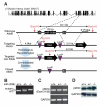
Figure 1. Distinct expression patterns of MyomiRs
A) Schematic representation of the genomic location of the miRNAs present in the different myosin genes. miR-208a and -208b share a comparable seed region (nt 2-8) but differ at three bases in their 3′ end (indicated in black). The seed region of miR-499 overlaps at 6 nt with miR-208 but differs significantly in the 3′ region. B) Northern analysis shows that miR-208a expression correlates with the cardiac-specific expression of α-MHC, while miR-208b parallels the predominant slow skeletal expression of β-MHC. miR-499 is co-expressed with Myh7b, a slow myosin that is mainly expressed in the heart and soleus, a slow skeletal muscle, but not in fast myofibers. Label indicates the mature miRNA. C) In situ hybridization shows that Myh7b is expressed specifically in the heart and somites as early as E12.5 during mouse embryogenesis. Silver grains are pseudocolored red. ht, heart; t, tongue.
Figure 2
Figure 2. miR-208a regulates Myh7b / miR-499
A) Northern blot analysis of heart and skeletal muscle tissue of miR-208a−/− animals shows that miR-499 expression is specifically extinguished in the heart while the expression in soleus remains unaffected. Compare with lower panel of Figure 1B. B) Northern blot analysis of heart tissue shows that miR-208a regulates the expression of myh7b/miR-499 in a stoichiometric manner. In miR-208a+/− animals, miR-499 and myh7b expression is reduced by 50%, while miR-499 and myh7b expression are eliminated in miR-208a−/− animals. Myh7b and GAPDH were detected by RT-PCR. GAPDH serves as a loading control. C) Northern blot analysis on cardiac samples of wild-type and miR-208a−/− (KO) neonate and adult mice. miR-208b and miR-499 are expressed normally in neonatal heart of miR-208a KO mice, whereas neither miRNA is expressed in adult miR-208a KO heart. D) Mice of the indicated miR-208a genotype were treated with PTU, as indicated, and miRNA expression was detected by Northern blot. PTU treatment increases miR-208b in wild-type animals, and this effect is significantly blunted in miR-208a−/− animals. E) Expression of miR-499 in hearts from wild type and MCK-miR-499 transgenic mice detected by Northern blot. F) Re-introducing miR-499 with an MCK-miR-499 transgene in the miR-208a−/− background restores expression of miR-208b and β-MHC in response to PTU treatment. Upper panels show Northern blots of hearts from duplicate animals under each condition. Lower panels show β-MHC expression as detected by real time RT-PCR. G) Fast skeletal muscle troponins (TnnT3 and TnnI2) are up-regulated in hearts of miR-208a−/− animals. Introduction of the MCK-miR-499 transgene into miR-208a−/− mice represses troponin expression as in wild-type. Results represent the average values obtained from two animals as detected by real time PCR.
Figure 3
Figure 3. Generation of miR-499 null mice
A) Strategy to generate miR-499 mutant mice by homologous recombination. The pre-miRNA sequence was replaced with a neomycin resistance cassette flanked by loxP sites. The neomycin cassette was removed in the mouse germline by breeding heterozygous mice to transgenic mice harboring the CAG-Cre transgene. B) Detection of the miR-499 mutation by PCR. Primers flanking the loxP site in intron 19 of the Myh7b gene generate PCR products as indicated. The mutant band in the PCR reaction is larger than the wild type band due to replacement of the miRNA with a larger loxP site. C) Detection of miR-499 and miR-208a transcripts by Northern analysis of hearts from wild-type, heterozygout and miR-499 mutant mice. D) Detection of Myh7b expression by RT-PCR of RNA from heart and soleus of mice of the indicated genotypes using primers flanking intron 19. Deletion of miR-499 does not disrupt cardiac expression of Myh7b, while there is a slight decrease in expression in soleus. E) Western analysis of α-MHC and β-MHC protein levels in hearts of neonatal mice of the indicated genotypes in the absence (left) and presence (right) of PTU. Two mice of each genotype were analyzed. Unlike miR-208, miR-499 is not required for up-regulation of β-MHC in response to PTU.
Figure 4
Figure 4. Generation of miR-208b null mice
A) Strategy to generate miR-208b mutant mice by homologous recombination. The pre-miRNA sequence was replaced with a neomycin resistance cassette flanked by loxP sites. The neomycin cassette was removed in the mouse germline by breeding heterozygous mice to transgenic mice harboring the CAG-Cre transgene. B) Detection of the miR-208b mutation by PCR. Primers flanking the loxP site in intron 31 of the β-MHC gene generate PCR products as indicated. C) Detection of β-MHC expression by RT-PCR of RNA from hearts of mice of the indicated genotypes using primers flanking intron 31. Deletion of miR-208b does not disrupt expression of β-MHC. D) Detection of β-MHC by western blot analysis of heart from P0 mice of the indicated genotypes, showing that β-MHC expression is not altered by deletion of miR-208b. GAPDH was detected as a loading control.
Figure 5
Figure 5. Control of skeletal muscle fiber type by miR-208b and miR-499
A) Detection of type I myofibers in soleus of wild-type, miR-208b−/−/miR-499−/− dKO, and MCK-miR-499 transgenic mice by metachromatic ATPase staining. Scale bar, 500 μm. B) Immunohistochemistry for βMHC to identify type I myofibers in the soleus of wild-type, miR-208b−/−/miR-499−/− dKO, and MCK-miR-499 transgenic mice. Scale bar, 500 μm. C) Identification of myosin isoform content of Soleus, EDL, and TA muscles from wild-type, miR-208b−/−/miR-499−/− dKO, and MCK-miR-499 transgenic mice by gel electrophoresis. D) The percentage of type I myofibers within soleus muscles of wild type, MyomiR mutant, and MCK-miR-499 transgenic mice was determined as shown in Panel B. E) βMHC mRNA levels in skeletal muscle of miR-208b−/−/miR-499−/− dKO adult mice were detected by real time PCR. Loss of MyomiRs in skeletal muscle results in repression of βMHC. F) Expression of slow and fast myofiber genes in soleus, TA and EDL muscles of MCK-miR-499 transgenic mice compared to their level of expression in wild type muscles (set at a value of 1), as detected by real time PCR. Transgenic over-expression of miR-499 is sufficient to up-regulate slow myofiber genes and repress fast myofiber genes in soleus and EDL. G) WT (n = 4) and MCK-miR-499 transgenic (n = 5) mice were subjected to a regimen of forced running to exhaustion on a treadmill. The time to run to exhaustion of each mouse is shown. p = 0.0039.
Figure 6
Figure 6. MyomiRs directly repress predicted target genes
A) Luciferase studies of predicted MyomiR target transcripts. Wild-type and mutant Sox6, Purβ, Sp3, and HP1β 3′UTRs were cloned into a luciferase reporter plasmid. These constructs were co-transfected into COS-1 cells with expression vectors for miR-208a or miR-499. Luminescence values were assayed forty-eight hours post transfection and were normalized to β-galactosidase activity. B) Expression of MyomiRs increases following C2C12 myoblast differentiation. Relative levels of miR-208b and miR-499 expression were detected by real-time PCR at days 0 and 5 of C2C12 differentiation. C) Wild-type, but not mutant luciferase reporter constructs are repressed by MyomiRs in C2C12 cells.
Figure 7
Figure 7. Target proteins that mediate the actions of MyomiRs
A) Western blot analysis of miR-208a/499 target expression in adult wild type (WT) and miR-208a−/− (KO) mouse heart lysate. Relative expression was calculated from comparison of band intensity following normalization to GAPDH loading control. B) Sox6 mRNA levels are elevated in the miR-208a−/− heart as measured by real time PCR. Transgenic expression of miR-499 abolished the increase in Sox6 mRNA in hearts lacking miR-208. C) Myh7b and miR-499 are repressed in the hearts of MCK-Sox6 transgenic mice. Transcript levels were measured in the hearts of P8 transgenic mice and wild type controls (WT) by real time PCR. D) Real time PCR indicates Sox6 mRNA levels are depressed in skeletal muscle of MCK-miR-499 mice. E) Sox6 mRNA transcript is overexpressed in MyomiR dKO skeletal muscle. MCK-Sox6 transgenic mice exhibit repression of slow skeletal muscle genes and unaltered levels of fast skeletal troponins in skeletal muscle. Expression was measured by real time PCR in total hind limb muscle of MCK-Sox6 transgenic mice and wild type (WT) controls. F) Immunohistochemistry for Type I myosin (βMHC) in the hind limb of MCK-Sox6 transgenic mice indicates an absence of Type I fibers in the EDL of transgenic mice as compared to wild-type (WT) littermate controls. Scale bar, 100 μm. G) Purβ mRNA transcript is overexpressed in MyomiR dKO skeletal muscle. MCK-Purβ transgenic mice demonstrate repression of slow skeletal muscle genes and unaltered fast skeletal gene expression. Expression was measured by real time PCR in total hind limb muscle of MCK-Purβ transgenic mice and wild type (WT) controls.
Figure 8
Figure 8. Control of myosin and fast versus slow muscle gene expression by the MyomiR network
In the adult heart, miR-208a encoded by the α-MHC gene is required for expression of β-MHC and Myh7b, which encode miR-208b and miR-499, respectively. Activation of Myh7b by miR-208a is constitutive, whereas activation of β-MHC also requires stress signals or absence of thyroid hormone. In slow skeletal muscle, Myh7b and β-MHC are regulated independently of miR-208a. miR-208a, -208b, and -499 repress the expression of a common set of transcriptional repressors that repress slow myosin and the slow myofiber gene program at the expense of fast muscle gene expression. Activation of the slow myofiber gene program also creates a positive feedback loop via the expression of miR-208b and miR-499, which further reinforce slow muscle gene program. Regulatory interactions within the blue box are shared by cardiac and slow skeletal muscle.

Similar articles

Cited by

References

    1. Adolph EA, Subramaniam A, Cserjesi P, Olson EN, Robbins J. Role of myocyte-specific enhancer-binding factor (MEF-2) in transcriptional regulation of the alpha-cardiac myosin heavy chain gene. J Biol Chem. 1993;268:5349–5352. - PubMed
    1. Azakie A, Fineman JR, He Y. Sp3 inhibits Sp1-mediated activation of the cardiac troponin T promoter and is downregulated during pathological cardiac hypertrophy in vivo. Am J Physiol Heart Circ Physiol. 2006;291:H600–611. - PubMed
    1. Baek D, Villen J, Shin C, Camargo FD, Gygi SP, Bartel DP. The impact of microRNAs on protein output. Nature. 2008;455:64–71. - PMC - PubMed
    1. Baldwin KM, Haddad F. Effects of different activity and inactivity paradigms on myosin heavy chain gene expression in striated muscle. J Appl Physiol. 2001;90:345–357. - PubMed
    1. Bartel DP. MicroRNAs: genomics, biogenesis, mechanism, and function. Cell. 2004;116:281–297. - PubMed

Publication types